


Hunter Schone
@hunterschone.bsky.social
www.hunterschone.com
Assistive technologies and neuroplasticity | NIH BRAIN Initiative postdoctoral fellow in the Collinger lab at the University of Pittsburgh | Prev: UCL and NIMHgov | 🏳️🌈
Assistive technologies and neuroplasticity | NIH BRAIN Initiative postdoctoral fellow in the Collinger lab at the University of Pittsburgh | Prev: UCL and NIMHgov | 🏳️🌈
We can only access the persistent ‘winner’ in humans because we can ask them to move their phantom fingers
We can perform this winner-takes-all analysis to replicate this supposed remapping, if we ignore the missing hand (red) and assign the territory to the next winner: lips (blue) or feet (green)
We can perform this winner-takes-all analysis to replicate this supposed remapping, if we ignore the missing hand (red) and assign the territory to the next winner: lips (blue) or feet (green)

August 21, 2025 at 8:35 PM
We can only access the persistent ‘winner’ in humans because we can ask them to move their phantom fingers
We can perform this winner-takes-all analysis to replicate this supposed remapping, if we ignore the missing hand (red) and assign the territory to the next winner: lips (blue) or feet (green)
We can perform this winner-takes-all analysis to replicate this supposed remapping, if we ignore the missing hand (red) and assign the territory to the next winner: lips (blue) or feet (green)
This does not mean that the monkeys’ brains forged new connections or that neurons changed their tuning because of the amputation, just that the region was already residually responsive to the adjacent fingers. You’re simply unmasking the adjacent digits, which you now assign to that territory.

August 21, 2025 at 8:35 PM
This does not mean that the monkeys’ brains forged new connections or that neurons changed their tuning because of the amputation, just that the region was already residually responsive to the adjacent fingers. You’re simply unmasking the adjacent digits, which you now assign to that territory.
They then assigned each cortical territory to the finger that elicited the greatest neuronal response when it was being touched, but because they could not touch the missing finger, the missing digit cannot be assigned any territory, because you can’t physically touch it.

August 21, 2025 at 8:35 PM
They then assigned each cortical territory to the finger that elicited the greatest neuronal response when it was being touched, but because they could not touch the missing finger, the missing digit cannot be assigned any territory, because you can’t physically touch it.
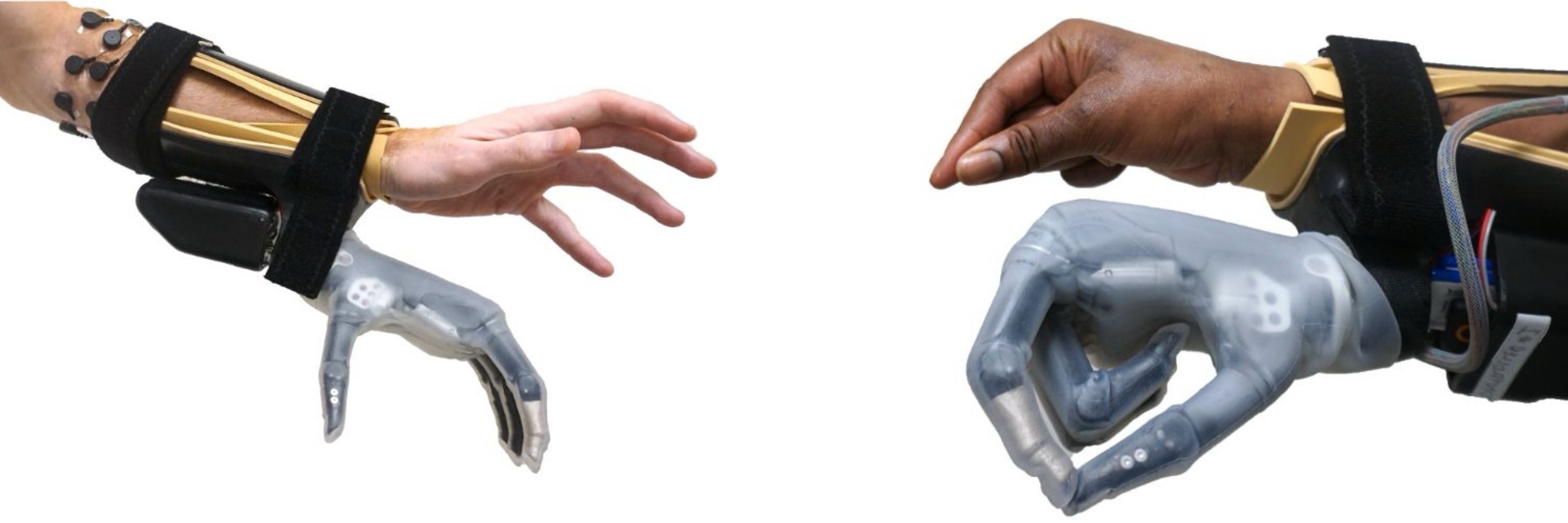
What do our results mean for brain-computer interfaces?
BCIs from #Neuralink and #BlackrockNeurotech work well for people with spinal cord injuries. Our findings suggest the sensory-deprived representations they tap into will remain stable despite long-term loss of a sensory input
🧵16/18
BCIs from #Neuralink and #BlackrockNeurotech work well for people with spinal cord injuries. Our findings suggest the sensory-deprived representations they tap into will remain stable despite long-term loss of a sensory input
🧵16/18
August 21, 2025 at 9:20 AM
What do our results mean for brain-computer interfaces?
BCIs from #Neuralink and #BlackrockNeurotech work well for people with spinal cord injuries. Our findings suggest the sensory-deprived representations they tap into will remain stable despite long-term loss of a sensory input
🧵16/18
BCIs from #Neuralink and #BlackrockNeurotech work well for people with spinal cord injuries. Our findings suggest the sensory-deprived representations they tap into will remain stable despite long-term loss of a sensory input
🧵16/18
While our results center on longitudinal data from 3 adults scanned before and after amputation, it’s supported by 4 fMRI control datasets from able-bodied and amputee participants. Combined, the study includes 166 scans across 86 people, the largest dataset devoted to cortical remapping
🧵14/18
🧵14/18

August 21, 2025 at 9:20 AM
While our results center on longitudinal data from 3 adults scanned before and after amputation, it’s supported by 4 fMRI control datasets from able-bodied and amputee participants. Combined, the study includes 166 scans across 86 people, the largest dataset devoted to cortical remapping
🧵14/18
🧵14/18
Perhaps, we might see changes if the patients were tested over a longer time period? To test this, we pooled data from chronic amputees (n=26) on average 23.5 years after amputation.
We see no unique differences in the hand or lip maps of our participants relative to this chronic group.
🧵13/18
We see no unique differences in the hand or lip maps of our participants relative to this chronic group.
🧵13/18

August 21, 2025 at 9:20 AM
Perhaps, we might see changes if the patients were tested over a longer time period? To test this, we pooled data from chronic amputees (n=26) on average 23.5 years after amputation.
We see no unique differences in the hand or lip maps of our participants relative to this chronic group.
🧵13/18
We see no unique differences in the hand or lip maps of our participants relative to this chronic group.
🧵13/18
So, the hand map is largely stable. But what about remapping of the adjacent body-part, the face?
From the lip data, we see the lips do not remap into the hand region. The patients show no unique changes vs able-bodied controls in magnitude or spread of lip activity in S1 post-amputation
🧵12/18
From the lip data, we see the lips do not remap into the hand region. The patients show no unique changes vs able-bodied controls in magnitude or spread of lip activity in S1 post-amputation
🧵12/18

August 21, 2025 at 9:20 AM
So, the hand map is largely stable. But what about remapping of the adjacent body-part, the face?
From the lip data, we see the lips do not remap into the hand region. The patients show no unique changes vs able-bodied controls in magnitude or spread of lip activity in S1 post-amputation
🧵12/18
From the lip data, we see the lips do not remap into the hand region. The patients show no unique changes vs able-bodied controls in magnitude or spread of lip activity in S1 post-amputation
🧵12/18
We trained a decoder on pre-amputation finger data and tested it on the phantom finger data. Across participants, decoding was significantly above chance, with slight decreases in finger-selective information over time.
🧵11/18
🧵11/18

August 21, 2025 at 9:20 AM
We trained a decoder on pre-amputation finger data and tested it on the phantom finger data. Across participants, decoding was significantly above chance, with slight decreases in finger-selective information over time.
🧵11/18
🧵11/18
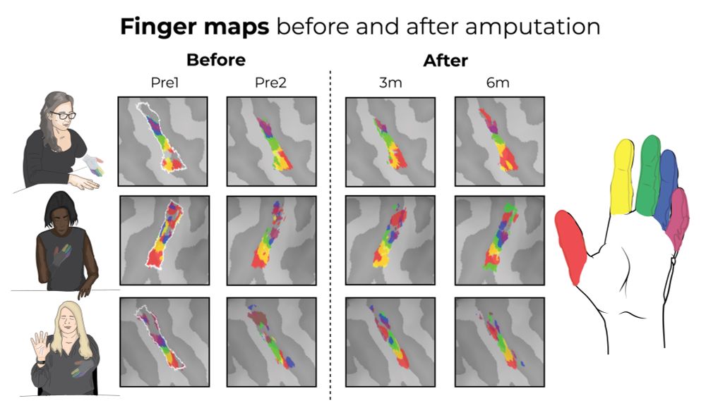
Similarly, when visualizing the selectivity maps for each finger before and after amputation, all participants show qualitatively similar maps before versus after amputation within S1
🧵10/18
🧵10/18
August 21, 2025 at 9:20 AM
Similarly, when visualizing the selectivity maps for each finger before and after amputation, all participants show qualitatively similar maps before versus after amputation within S1
🧵10/18
🧵10/18
Testing for changes at the individual voxel level in the S1 hand region, we performed a multivariate analysis correlating the activation pattern of each affected finger to the same phantom finger; this revealed the patterns are highly similar before and after amputation
🧵9/18
🧵9/18

August 21, 2025 at 9:20 AM
Testing for changes at the individual voxel level in the S1 hand region, we performed a multivariate analysis correlating the activation pattern of each affected finger to the same phantom finger; this revealed the patterns are highly similar before and after amputation
🧵9/18
🧵9/18
Focusing on somatosensory cortex (S1: the region receiving the altered sensory input), we investigated whether there are finer grained changes happening. We see the pre-amputated fingers and phantom fingers have the same magnitude and spread of activity across S1.
🧵8/18
🧵8/18

August 21, 2025 at 9:20 AM
Focusing on somatosensory cortex (S1: the region receiving the altered sensory input), we investigated whether there are finer grained changes happening. We see the pre-amputated fingers and phantom fingers have the same magnitude and spread of activity across S1.
🧵8/18
🧵8/18
Qualitatively, the maps for the hand (before) and phantom hand (after) and lips (before/after) are strikingly stable across the amputation for all participants!
🧵7/18
🧵7/18

August 21, 2025 at 9:20 AM
Qualitatively, the maps for the hand (before) and phantom hand (after) and lips (before/after) are strikingly stable across the amputation for all participants!
🧵7/18
🧵7/18
During MRI scanning, participants tapped individual fingers, pursed their lips and curled their toes. The same task was performed before and after amputation. Post-amputation, participants attempted to perform the same task with their phantom fingers.
🧵6/18
🧵6/18

August 21, 2025 at 9:20 AM
During MRI scanning, participants tapped individual fingers, pursed their lips and curled their toes. The same task was performed before and after amputation. Post-amputation, participants attempted to perform the same task with their phantom fingers.
🧵6/18
🧵6/18
Using 3T functional MRI @imagingneuroucl.bsky.social we scanned patients twice before the amputation surgery and 3 months, 6 months, 1.5 years and even up to 5 years after the amputation.
Also, 16 able-bodied controls were scanned 4 times, over 6 months.
🧵5/18
Also, 16 able-bodied controls were scanned 4 times, over 6 months.
🧵5/18

August 21, 2025 at 9:20 AM
Using 3T functional MRI @imagingneuroucl.bsky.social we scanned patients twice before the amputation surgery and 3 months, 6 months, 1.5 years and even up to 5 years after the amputation.
Also, 16 able-bodied controls were scanned 4 times, over 6 months.
🧵5/18
Also, 16 able-bodied controls were scanned 4 times, over 6 months.
🧵5/18
After amputation, all patients reported vivid phantom sensations. Crucially, moving a phantom limb is not imaginary. They physically attempt the movements, shown by residual limb contractions and selective sensorimotor activity for attempted vs imagined phantom movements
🧵4/18
🧵4/18
August 21, 2025 at 9:20 AM
After amputation, all patients reported vivid phantom sensations. Crucially, moving a phantom limb is not imaginary. They physically attempt the movements, shown by residual limb contractions and selective sensorimotor activity for attempted vs imagined phantom movements
🧵4/18
🧵4/18
We aimed to longitudinally track the brain’s body map in patients with planned arm amputations, before and after surgery. Over 8 years across multiple NHS sites, we tested 3 patients with planned arm amputations, for reasons including vascular malformation and cancer.
🧵3/18
🧵3/18
August 21, 2025 at 9:20 AM
We aimed to longitudinally track the brain’s body map in patients with planned arm amputations, before and after surgery. Over 8 years across multiple NHS sites, we tested 3 patients with planned arm amputations, for reasons including vascular malformation and cancer.
🧵3/18
🧵3/18
Early research on monkey amputations argued for dramatic somatosensory cortex remapping after amputation. But these, and similar human studies, used cross-sectional study designs, not directly tracking changes over time in the same monkey/human… that is until now.
🧵2/18
🧵2/18

August 21, 2025 at 9:20 AM
Early research on monkey amputations argued for dramatic somatosensory cortex remapping after amputation. But these, and similar human studies, used cross-sectional study designs, not directly tracking changes over time in the same monkey/human… that is until now.
🧵2/18
🧵2/18
Now out in @natneuro.nature.com
What happens to the brain’s body map when a body-part is removed?
Scanning patients before and up to 5 yrs after arm amputation, we discovered the brain’s body map is strikingly preserved despite amputation
www.nature.com/articles/s41593-025-02037-7
🧵1/18
What happens to the brain’s body map when a body-part is removed?
Scanning patients before and up to 5 yrs after arm amputation, we discovered the brain’s body map is strikingly preserved despite amputation
www.nature.com/articles/s41593-025-02037-7
🧵1/18
August 21, 2025 at 9:20 AM
Now out in @natneuro.nature.com
What happens to the brain’s body map when a body-part is removed?
Scanning patients before and up to 5 yrs after arm amputation, we discovered the brain’s body map is strikingly preserved despite amputation
www.nature.com/articles/s41593-025-02037-7
🧵1/18
What happens to the brain’s body map when a body-part is removed?
Scanning patients before and up to 5 yrs after arm amputation, we discovered the brain’s body map is strikingly preserved despite amputation
www.nature.com/articles/s41593-025-02037-7
🧵1/18
@standupforscience.bsky.social in Pittsburgh with @jeffweiss.bsky.social and Brian Dekleva


March 7, 2025 at 6:05 PM
@standupforscience.bsky.social in Pittsburgh with @jeffweiss.bsky.social and Brian Dekleva