


Parichy Lab | Dave Parichy
@dparichy.bsky.social
developmental genetics, genomics of adult traits and evolution | zebrafish, danios, guppy, wrasses, mandarin fish | neural crest, stem cells, pigment, skin | University of Virginia Biology | parichylab.org | #DevBio #EvoDevo #NeuralCrest
Like other dominant pigment pattern mutants, this allele is named for a surrealist, in this case the Spanish-Mexican painter Remedios Varo. Here is one of her works, Allegory of Winter (1948), which as a bonus seems to show dendrites extending from the "cells."

March 24, 2025 at 2:28 PM
Like other dominant pigment pattern mutants, this allele is named for a surrealist, in this case the Spanish-Mexican painter Remedios Varo. Here is one of her works, Allegory of Winter (1948), which as a bonus seems to show dendrites extending from the "cells."
These and other genetic analyses in zebrafish, as well as studies of mutants in other species, suggest a model for evolution of Mitf requirements in pigment cell lineages.

March 24, 2025 at 2:23 PM
These and other genetic analyses in zebrafish, as well as studies of mutants in other species, suggest a model for evolution of Mitf requirements in pigment cell lineages.
This phenotype is recapitulated when the Mitf null allele is combined with a null allele for Tfec.

March 24, 2025 at 2:21 PM
This phenotype is recapitulated when the Mitf null allele is combined with a null allele for Tfec.
In both species the allele is semi-dominant due to mutation in DNA binding domain. Although recessive alleles of Mitf in zebrafish have melanophore defects, we found that this allele also has defects in xanthophores, too.

March 24, 2025 at 2:16 PM
In both species the allele is semi-dominant due to mutation in DNA binding domain. Although recessive alleles of Mitf in zebrafish have melanophore defects, we found that this allele also has defects in xanthophores, too.
With forward genetic screen, found exact same mutation in fish as identified many years ago in mouse.

March 24, 2025 at 2:13 PM
With forward genetic screen, found exact same mutation in fish as identified many years ago in mouse.
For sure… did my best to make that clear!
February 22, 2025 at 4:16 PM
For sure… did my best to make that clear!
“Yeah what about it” was pretty much exactly what McGuire(R) VA-5 staffer had to say when contacted
February 22, 2025 at 3:18 AM
“Yeah what about it” was pretty much exactly what McGuire(R) VA-5 staffer had to say when contacted
VA-5 (R) staffer: nothing to be done, government just can’t afford these costs anymore.
February 22, 2025 at 3:12 AM
VA-5 (R) staffer: nothing to be done, government just can’t afford these costs anymore.
As usual, a heroic job from our own folks, especially Dylan, and great fun working with our collaborators, and of course thanks to NIH and the MIRA mechanism for letting us go where the science leads.
February 18, 2025 at 9:07 PM
As usual, a heroic job from our own folks, especially Dylan, and great fun working with our collaborators, and of course thanks to NIH and the MIRA mechanism for letting us go where the science leads.
We believe this is all pretty cool because (i) it shows how a differentiated cell type has been “repurposed” to have an entirely new phenotype and (ii) it shows the importance of positional information even in a species best known for self-organizing its pattern.
February 18, 2025 at 9:07 PM
We believe this is all pretty cool because (i) it shows how a differentiated cell type has been “repurposed” to have an entirely new phenotype and (ii) it shows the importance of positional information even in a species best known for self-organizing its pattern.
Putting it all together, we think this is what’s going on.
February 18, 2025 at 9:04 PM
Putting it all together, we think this is what’s going on.
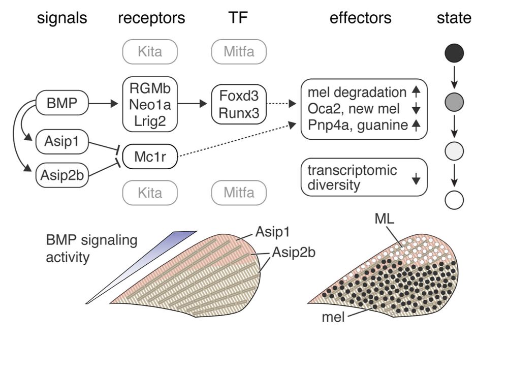
So how do BMP (and Agouti) drive fate conversion? Transcription factors Foxd3 (well known) and Runx3 (not well known in pigment biology) are downstream of BMP and necessary and sufficient to effect melanin loss and purine gain.
February 18, 2025 at 9:02 PM
So how do BMP (and Agouti) drive fate conversion? Transcription factors Foxd3 (well known) and Runx3 (not well known in pigment biology) are downstream of BMP and necessary and sufficient to effect melanin loss and purine gain.
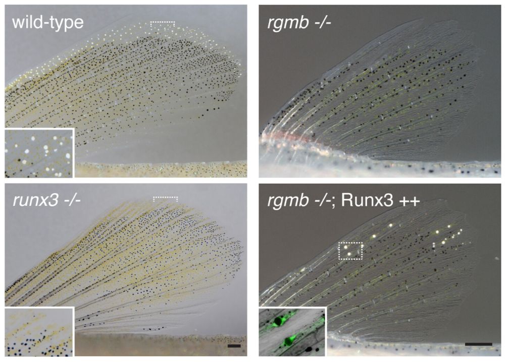
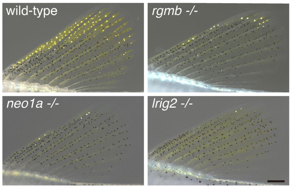
But even knocking out both Agouti genes didn’t eliminate all the white cells so we looked further and identified an additional role for BMPs, which pattern the fin more generally. Here BMPs signal through non-canonical receptors including Rgmb.
February 18, 2025 at 9:02 PM
But even knocking out both Agouti genes didn’t eliminate all the white cells so we looked further and identified an additional role for BMPs, which pattern the fin more generally. Here BMPs signal through non-canonical receptors including Rgmb.
We hypothesized that positional information specifies where transdifferentiation should happen. scRNA-seq, mutants and transgenes showed that one such cue is Agouti-related Asip1, which acts in concert with another Agouti peptide, Asip2b,

February 18, 2025 at 9:01 PM
We hypothesized that positional information specifies where transdifferentiation should happen. scRNA-seq, mutants and transgenes showed that one such cue is Agouti-related Asip1, which acts in concert with another Agouti peptide, Asip2b,
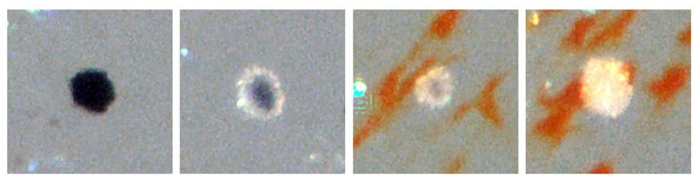
Here’s a kickass FIB-SEM movie of a transitional cell, showing organelles in various states of degradation or development (this took a week of instrument time).
February 18, 2025 at 9:01 PM
Here’s a kickass FIB-SEM movie of a transitional cell, showing organelles in various states of degradation or development (this took a week of instrument time).
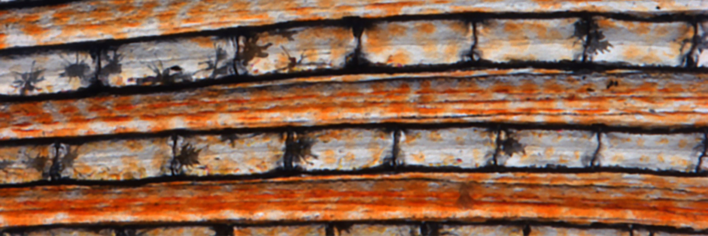
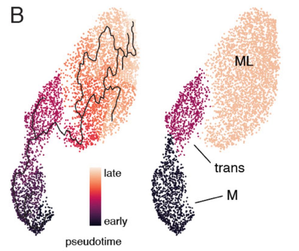
We showed previously that white cells—melanoleucophores—differentiate from melanophores. Here we show how this occurs, with massive changes in transcriptome, turnover of pigmentary organelles, loss of melanin and gain of white guanine crystals.
February 18, 2025 at 9:00 PM
We showed previously that white cells—melanoleucophores—differentiate from melanophores. Here we show how this occurs, with massive changes in transcriptome, turnover of pigmentary organelles, loss of melanin and gain of white guanine crystals.
Ontogenetic, nuptial, physiological? Several possible contacts.
January 26, 2025 at 8:52 PM
Ontogenetic, nuptial, physiological? Several possible contacts.