


Dominik Robak
@dominikrobak.bsky.social
Computers and microscopes are cool
Happy #MicroscopyMonday!
Bees didn’t invent the honeycomb—biology’s been doing it at the single-cell level all along.
🟢 GFP-tagged junctions
🔵 Hoechst-stained nuclei
A living mouse intestinal villus.
No blueprint. Just (bio)physics.
#intravitalimaging #biophysics #OME #microscopyenvironment
Bees didn’t invent the honeycomb—biology’s been doing it at the single-cell level all along.
🟢 GFP-tagged junctions
🔵 Hoechst-stained nuclei
A living mouse intestinal villus.
No blueprint. Just (bio)physics.
#intravitalimaging #biophysics #OME #microscopyenvironment

April 22, 2025 at 12:35 AM
Happy #MicroscopyMonday!
Bees didn’t invent the honeycomb—biology’s been doing it at the single-cell level all along.
🟢 GFP-tagged junctions
🔵 Hoechst-stained nuclei
A living mouse intestinal villus.
No blueprint. Just (bio)physics.
#intravitalimaging #biophysics #OME #microscopyenvironment
Bees didn’t invent the honeycomb—biology’s been doing it at the single-cell level all along.
🟢 GFP-tagged junctions
🔵 Hoechst-stained nuclei
A living mouse intestinal villus.
No blueprint. Just (bio)physics.
#intravitalimaging #biophysics #OME #microscopyenvironment
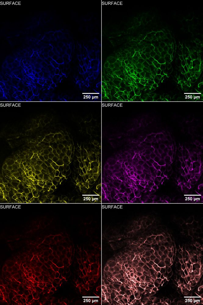
Bonus: ran a quick color variation test — because it's always worth playing with presentation.
Color isn’t just style — it shapes what you see.
#MicroscopyCommunity #ScientificVisualization #Biophysics
Color isn’t just style — it shapes what you see.
#MicroscopyCommunity #ScientificVisualization #Biophysics
April 14, 2025 at 7:01 PM
Bonus: ran a quick color variation test — because it's always worth playing with presentation.
Color isn’t just style — it shapes what you see.
#MicroscopyCommunity #ScientificVisualization #Biophysics
Color isn’t just style — it shapes what you see.
#MicroscopyCommunity #ScientificVisualization #Biophysics
#MicroscopyMonday | Colon-scoping turned coral reef
Was imaging the colon when I accidentally stumbled into this glowing patch of vascularized white adipose tissue.
🔵 mT/mG mouse (mT-only) → red membranes shown in cyan
Accidental imaging hits different.
#IntravitalImaging #LiveImaging
Was imaging the colon when I accidentally stumbled into this glowing patch of vascularized white adipose tissue.
🔵 mT/mG mouse (mT-only) → red membranes shown in cyan
Accidental imaging hits different.
#IntravitalImaging #LiveImaging


April 14, 2025 at 7:01 PM
#MicroscopyMonday | Colon-scoping turned coral reef
Was imaging the colon when I accidentally stumbled into this glowing patch of vascularized white adipose tissue.
🔵 mT/mG mouse (mT-only) → red membranes shown in cyan
Accidental imaging hits different.
#IntravitalImaging #LiveImaging
Was imaging the colon when I accidentally stumbled into this glowing patch of vascularized white adipose tissue.
🔵 mT/mG mouse (mT-only) → red membranes shown in cyan
Accidental imaging hits different.
#IntravitalImaging #LiveImaging
#3DThursday | Intestinal villi in all their folded glory
These self-renewing, nutrient-absorbing structures are beautiful and functional.
🔴 Phalloidin
🔵 DAPI
🟢 Clvd cas-3 (extrusion in action!)
Fixed tissue 3D imaging = big insight, tiny resolution.
#Microscopy #3DImaging #biophysics #intestine
These self-renewing, nutrient-absorbing structures are beautiful and functional.
🔴 Phalloidin
🔵 DAPI
🟢 Clvd cas-3 (extrusion in action!)
Fixed tissue 3D imaging = big insight, tiny resolution.
#Microscopy #3DImaging #biophysics #intestine
April 11, 2025 at 3:08 AM
#3DThursday | Intestinal villi in all their folded glory
These self-renewing, nutrient-absorbing structures are beautiful and functional.
🔴 Phalloidin
🔵 DAPI
🟢 Clvd cas-3 (extrusion in action!)
Fixed tissue 3D imaging = big insight, tiny resolution.
#Microscopy #3DImaging #biophysics #intestine
These self-renewing, nutrient-absorbing structures are beautiful and functional.
🔴 Phalloidin
🔵 DAPI
🟢 Clvd cas-3 (extrusion in action!)
Fixed tissue 3D imaging = big insight, tiny resolution.
#Microscopy #3DImaging #biophysics #intestine
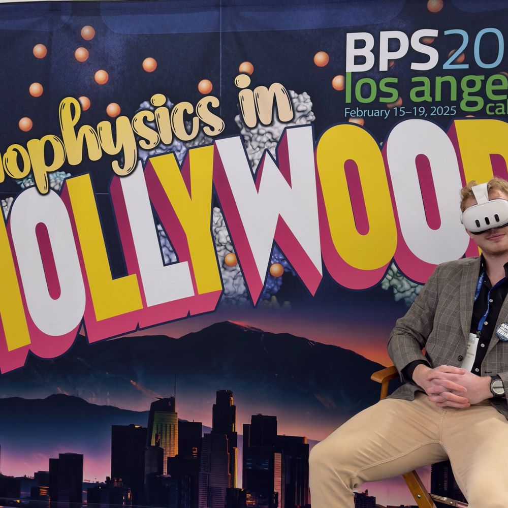
Hey #BPS2025! Wanna look at cool pictures and see what happens in mice’s intestine throughout live imaging? Come by my poster at LB69 on Monday to learn more! Got some cool stuff to experience imaging data like never before! #Biophysics #Science #Microscopy #VR

February 17, 2025 at 7:58 AM
Hey #BPS2025! Wanna look at cool pictures and see what happens in mice’s intestine throughout live imaging? Come by my poster at LB69 on Monday to learn more! Got some cool stuff to experience imaging data like never before! #Biophysics #Science #Microscopy #VR