


Denis Krndija
@deniskrndija.bsky.social
new group leader @CBIToulouse | MechanoGut team | ATIP-Avenir | fascinated by the dynamics and mechanics of the #gut 🐭⚙️🔬| he/his/him 🏳️🌈
Lab: https://cbi-toulouse.fr/eng/equipe-krndija
Personal: https://cbi-toulouse.fr/eng/page-personnelle-49
Lab: https://cbi-toulouse.fr/eng/equipe-krndija
Personal: https://cbi-toulouse.fr/eng/page-personnelle-49
Using a luminal-accessibility assay for E-cadherin, we found that depleting NMIIA in vivo or inhibiting myosin II / Ca²⁺ influx ex vivo led to barrier breaches under mechanical stress.
The junctional response is therefore essential for maintaining epithelial integrity (10/11)
The junctional response is therefore essential for maintaining epithelial integrity (10/11)

October 8, 2025 at 10:17 PM
Using a luminal-accessibility assay for E-cadherin, we found that depleting NMIIA in vivo or inhibiting myosin II / Ca²⁺ influx ex vivo led to barrier breaches under mechanical stress.
The junctional response is therefore essential for maintaining epithelial integrity (10/11)
The junctional response is therefore essential for maintaining epithelial integrity (10/11)
Conversely, activating Piezo1 was enough to trigger NMII activation and junctional recruitment.
👉 Mechanosensitive Ca²⁺ influx is both necessary and sufficient for junctional reinforcement under force ⚡️(9/11)
👉 Mechanosensitive Ca²⁺ influx is both necessary and sufficient for junctional reinforcement under force ⚡️(9/11)

October 8, 2025 at 10:17 PM
Conversely, activating Piezo1 was enough to trigger NMII activation and junctional recruitment.
👉 Mechanosensitive Ca²⁺ influx is both necessary and sufficient for junctional reinforcement under force ⚡️(9/11)
👉 Mechanosensitive Ca²⁺ influx is both necessary and sufficient for junctional reinforcement under force ⚡️(9/11)
💡 What activates myosin II under mechanical stress?
Blocking mechanosensitive ion channels (with Gd³⁺) or chelating extracellular Ca²⁺ (with BAPTA) prevented NMII activation and junctional reinforcement – stopping the mechano-adaptive response in its tracks (8/11)
Blocking mechanosensitive ion channels (with Gd³⁺) or chelating extracellular Ca²⁺ (with BAPTA) prevented NMII activation and junctional reinforcement – stopping the mechano-adaptive response in its tracks (8/11)

October 8, 2025 at 10:17 PM
💡 What activates myosin II under mechanical stress?
Blocking mechanosensitive ion channels (with Gd³⁺) or chelating extracellular Ca²⁺ (with BAPTA) prevented NMII activation and junctional reinforcement – stopping the mechano-adaptive response in its tracks (8/11)
Blocking mechanosensitive ion channels (with Gd³⁺) or chelating extracellular Ca²⁺ (with BAPTA) prevented NMII activation and junctional reinforcement – stopping the mechano-adaptive response in its tracks (8/11)
Genetic deletion (Myh9-KO in vivo) or pharmacological inhibition of NMII ex vivo abolished junctional recruitment across all complexes.
👉 NMII acts as a central effector coordinating force sensing and junctional reinforcement.🔩 (7/11)
👉 NMII acts as a central effector coordinating force sensing and junctional reinforcement.🔩 (7/11)

October 8, 2025 at 10:17 PM
Genetic deletion (Myh9-KO in vivo) or pharmacological inhibition of NMII ex vivo abolished junctional recruitment across all complexes.
👉 NMII acts as a central effector coordinating force sensing and junctional reinforcement.🔩 (7/11)
👉 NMII acts as a central effector coordinating force sensing and junctional reinforcement.🔩 (7/11)
Junctional reinforcement coincided with recruitment of myosin IIA (NMIIA) to perijunctional belts and transient apical constriction – hallmarks of contractile activation.
Unexpectedly, myosin IIC, usually linked to microvilli, also relocalized to junctions under force ⚙️ (6/11)
Unexpectedly, myosin IIC, usually linked to microvilli, also relocalized to junctions under force ⚙️ (6/11)

October 8, 2025 at 10:17 PM
Junctional reinforcement coincided with recruitment of myosin IIA (NMIIA) to perijunctional belts and transient apical constriction – hallmarks of contractile activation.
Unexpectedly, myosin IIC, usually linked to microvilli, also relocalized to junctions under force ⚙️ (6/11)
Unexpectedly, myosin IIC, usually linked to microvilli, also relocalized to junctions under force ⚙️ (6/11)
Using a controlled in vivo colonic distension system (with Nicolas Cenac, IRSD Toulouse), we uncovered two kinetic modes:
- Tight & adherens junctions → sustained reinforcement
- Desmosomes & keratin filaments → progressive accumulation over time (5/11)
- Tight & adherens junctions → sustained reinforcement
- Desmosomes & keratin filaments → progressive accumulation over time (5/11)

October 8, 2025 at 10:17 PM
Using a controlled in vivo colonic distension system (with Nicolas Cenac, IRSD Toulouse), we uncovered two kinetic modes:
- Tight & adherens junctions → sustained reinforcement
- Desmosomes & keratin filaments → progressive accumulation over time (5/11)
- Tight & adherens junctions → sustained reinforcement
- Desmosomes & keratin filaments → progressive accumulation over time (5/11)
This mucosal remodelling comes with striking reinforcement of tight, adherens, and desmosomal junctions – a robust, pan-junctional mechano-adaptive response.
The adult gut epithelium actively adapts to physiological mechanical stress 💪 (4/11)
The adult gut epithelium actively adapts to physiological mechanical stress 💪 (4/11)

October 8, 2025 at 10:17 PM
This mucosal remodelling comes with striking reinforcement of tight, adherens, and desmosomal junctions – a robust, pan-junctional mechano-adaptive response.
The adult gut epithelium actively adapts to physiological mechanical stress 💪 (4/11)
The adult gut epithelium actively adapts to physiological mechanical stress 💪 (4/11)
💩 Faeces matter – mechanically!
Most studies remove luminal contents before analysis – we didn’t.
Keeping them revealed that the colonic mucosa adapts to faecal distension through large-scale tissue unfolding and epithelial deformation (3/11)
Most studies remove luminal contents before analysis – we didn’t.
Keeping them revealed that the colonic mucosa adapts to faecal distension through large-scale tissue unfolding and epithelial deformation (3/11)

October 8, 2025 at 10:17 PM
💩 Faeces matter – mechanically!
Most studies remove luminal contents before analysis – we didn’t.
Keeping them revealed that the colonic mucosa adapts to faecal distension through large-scale tissue unfolding and epithelial deformation (3/11)
Most studies remove luminal contents before analysis – we didn’t.
Keeping them revealed that the colonic mucosa adapts to faecal distension through large-scale tissue unfolding and epithelial deformation (3/11)
Enjoying great science and stunning alpine views at the GRC on Cell Contact & Adhesion in Les Diablerets 🇨🇭⛰️ Proud of our PhD students Dhriti and Vishnu for their terrific work - posters and talks full of exciting insights! #CellContactAndAdhesionGRC #CellBiology #PhDLife #LesDiablerets

June 10, 2025 at 9:32 PM
Enjoying great science and stunning alpine views at the GRC on Cell Contact & Adhesion in Les Diablerets 🇨🇭⛰️ Proud of our PhD students Dhriti and Vishnu for their terrific work - posters and talks full of exciting insights! #CellContactAndAdhesionGRC #CellBiology #PhDLife #LesDiablerets
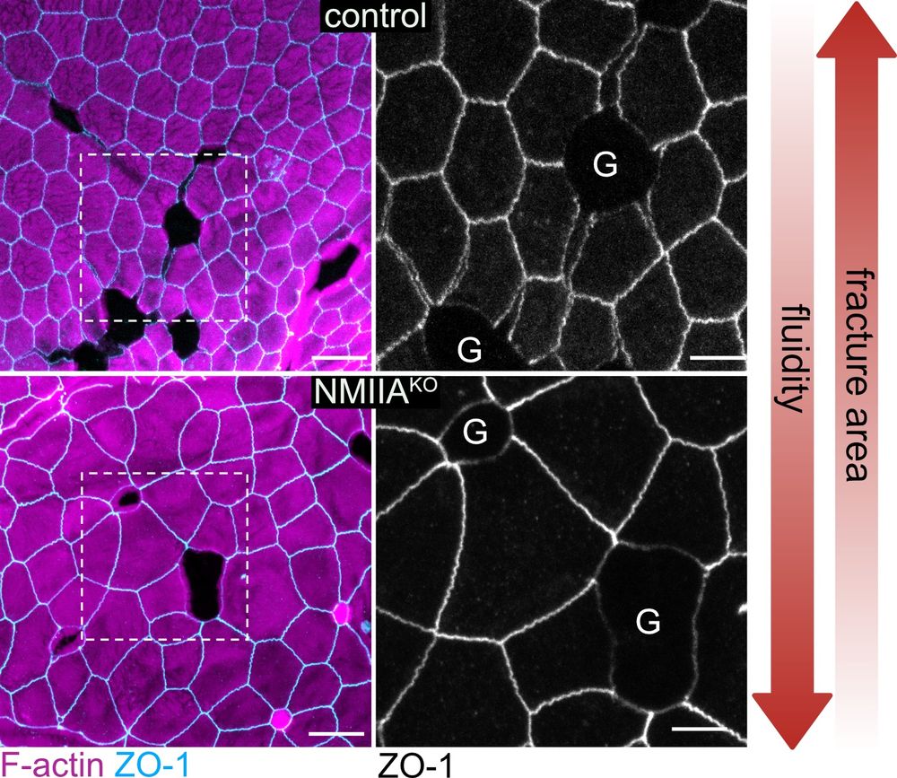
We tested our model predictions by modulating tissue rigidity via myosin II in vivo (NMIIA-KO) and in 2D organoids – tissue fluidification reduced GAFs, even under goblet cell hypertrophy! → It’s the balance between goblet pressure and tissue fluidity that controls junctional integrity! (8/9)
April 6, 2025 at 10:24 PM
We tested our model predictions by modulating tissue rigidity via myosin II in vivo (NMIIA-KO) and in 2D organoids – tissue fluidification reduced GAFs, even under goblet cell hypertrophy! → It’s the balance between goblet pressure and tissue fluidity that controls junctional integrity! (8/9)
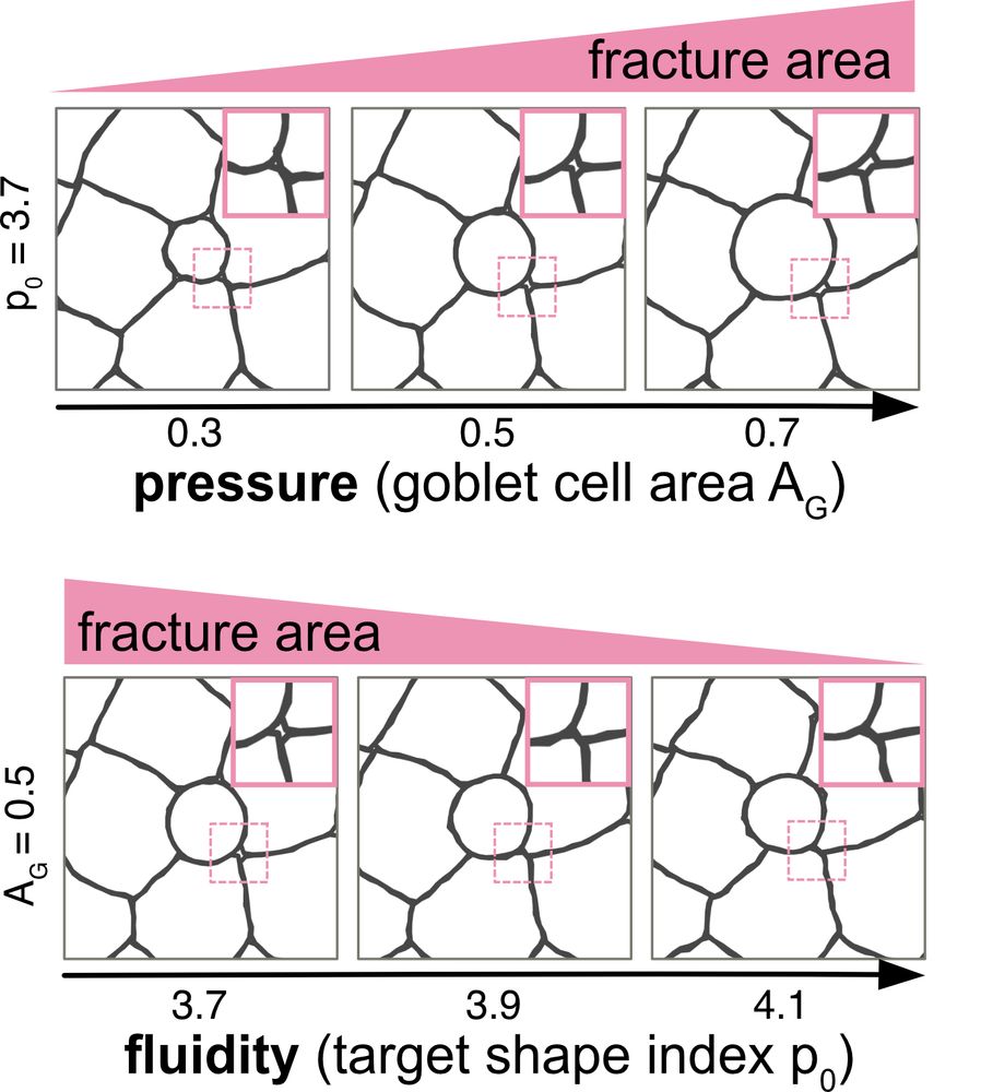
To understand the force balance behind rupture, we teamed up with Shi-Lei Xue 🖥️ (another new PI!👋). Through an innovative vertex model incorporating fractures, we identified 2 key GAF parameters:
1) Goblet size/pressure → bigger goblets = more GAFs
2) Tissue fluidity → more fluid = fewer GAFs! (7/9)
1) Goblet size/pressure → bigger goblets = more GAFs
2) Tissue fluidity → more fluid = fewer GAFs! (7/9)
April 6, 2025 at 10:24 PM
To understand the force balance behind rupture, we teamed up with Shi-Lei Xue 🖥️ (another new PI!👋). Through an innovative vertex model incorporating fractures, we identified 2 key GAF parameters:
1) Goblet size/pressure → bigger goblets = more GAFs
2) Tissue fluidity → more fluid = fewer GAFs! (7/9)
1) Goblet size/pressure → bigger goblets = more GAFs
2) Tissue fluidity → more fluid = fewer GAFs! (7/9)
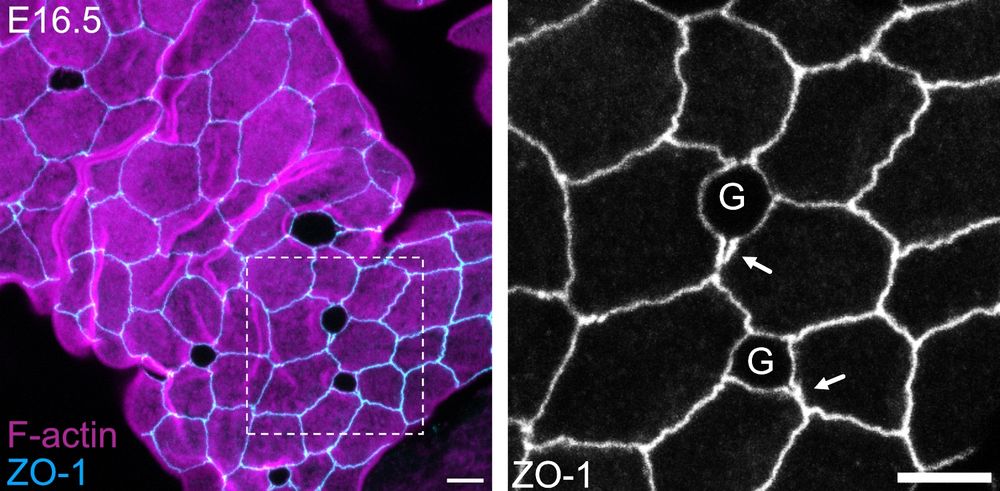
Are GAFs linked to food transit or microbiota? 🤔 Surprisingly, GAFs exist in embryonic intestine (E16.5), where the lumen is empty & neither microbiota nor immune cells are present! Plus, we reproduced GAFs in 2D organoids, confirming their epithelium-autonomous nature! 💡 (6/9)
April 6, 2025 at 10:24 PM
Are GAFs linked to food transit or microbiota? 🤔 Surprisingly, GAFs exist in embryonic intestine (E16.5), where the lumen is empty & neither microbiota nor immune cells are present! Plus, we reproduced GAFs in 2D organoids, confirming their epithelium-autonomous nature! 💡 (6/9)
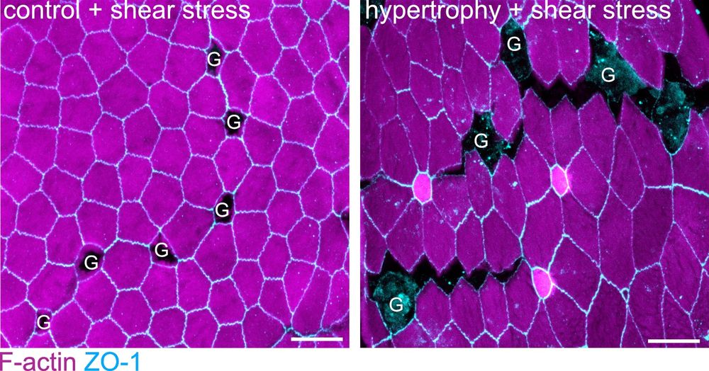
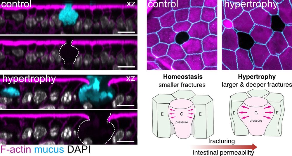
What about the extrinsic mechanical stresses in the gut? Goblet hypertrophy not only increases GAF size & number but also fracture depth (reaching desmosomes), weakening the epithelium’s resistance to luminal shear stress. This escalated into large-scale epithelial cracking in our exp.! 💥💥💥 🤯 (5/9)
April 6, 2025 at 10:24 PM
What about the extrinsic mechanical stresses in the gut? Goblet hypertrophy not only increases GAF size & number but also fracture depth (reaching desmosomes), weakening the epithelium’s resistance to luminal shear stress. This escalated into large-scale epithelial cracking in our exp.! 💥💥💥 🤯 (5/9)
Are enterocytes just passive observers?
No! They accumulate E-cadherin & other junctional proteins in a mechano-sensitive response to goblet-induced strain! Yet, goblet hypertrophy overcomes this active response, leading to full junctional failure in a tight > adherens > desmosome hierarchy! ⚠️ (4/9)
No! They accumulate E-cadherin & other junctional proteins in a mechano-sensitive response to goblet-induced strain! Yet, goblet hypertrophy overcomes this active response, leading to full junctional failure in a tight > adherens > desmosome hierarchy! ⚠️ (4/9)
April 6, 2025 at 10:24 PM
Are enterocytes just passive observers?
No! They accumulate E-cadherin & other junctional proteins in a mechano-sensitive response to goblet-induced strain! Yet, goblet hypertrophy overcomes this active response, leading to full junctional failure in a tight > adherens > desmosome hierarchy! ⚠️ (4/9)
No! They accumulate E-cadherin & other junctional proteins in a mechano-sensitive response to goblet-induced strain! Yet, goblet hypertrophy overcomes this active response, leading to full junctional failure in a tight > adherens > desmosome hierarchy! ⚠️ (4/9)
How do goblets induce GAFs?
They compress & deform neighboring enterocytes, straining their junctions to the point of rupture! GAFs are regulated by goblet cell volume – goblet hypertrophy, often a protective response (parasite clearance), paradoxically increases fracturing & permeability! 🤯 (3/9)
They compress & deform neighboring enterocytes, straining their junctions to the point of rupture! GAFs are regulated by goblet cell volume – goblet hypertrophy, often a protective response (parasite clearance), paradoxically increases fracturing & permeability! 🤯 (3/9)
April 6, 2025 at 10:24 PM
How do goblets induce GAFs?
They compress & deform neighboring enterocytes, straining their junctions to the point of rupture! GAFs are regulated by goblet cell volume – goblet hypertrophy, often a protective response (parasite clearance), paradoxically increases fracturing & permeability! 🤯 (3/9)
They compress & deform neighboring enterocytes, straining their junctions to the point of rupture! GAFs are regulated by goblet cell volume – goblet hypertrophy, often a protective response (parasite clearance), paradoxically increases fracturing & permeability! 🤯 (3/9)
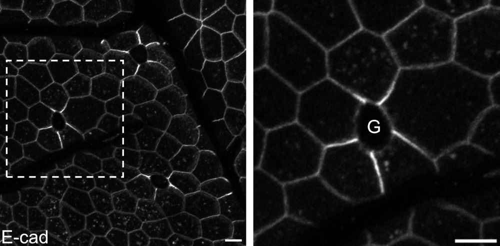
We identified Goblet cell-Associated Fractures (GAFs) 💥 – junctional ruptures between goblet-adjacent enterocytes in normal adult mouse and human intestine! To our knowledge, this is the first example of a self-rupturing tissue under homeostatic conditions! 🧐 (2/9)

April 6, 2025 at 10:24 PM
We identified Goblet cell-Associated Fractures (GAFs) 💥 – junctional ruptures between goblet-adjacent enterocytes in normal adult mouse and human intestine! To our knowledge, this is the first example of a self-rupturing tissue under homeostatic conditions! 🧐 (2/9)
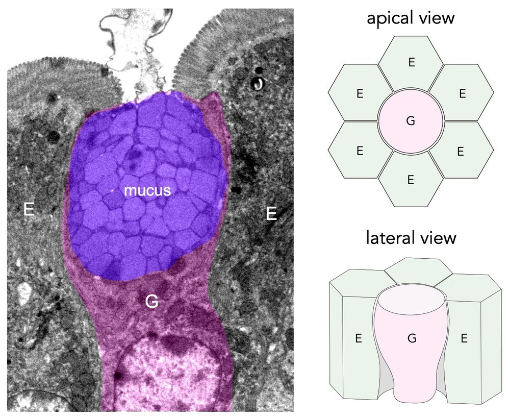
🥳 Thrilled to share our lab's first preprint, led by our talented postdoc Justine Creff! 👩🏻🔬 We tackled a fundamental question: how the epithelium withstands mechanical stress at the interface of cells with distinct geometries and mechanics, such as enterocytes (E) and goblet cells (G) (1/9)
April 6, 2025 at 10:24 PM
🥳 Thrilled to share our lab's first preprint, led by our talented postdoc Justine Creff! 👩🏻🔬 We tackled a fundamental question: how the epithelium withstands mechanical stress at the interface of cells with distinct geometries and mechanics, such as enterocytes (E) and goblet cells (G) (1/9)
Happy holidays 🎄 and #FluorescenceFriday with this festive mouse colonoid! Work of Amalia Ferran, new PhD student in our team @cbitoulouse.bsky.social 🙏🐭🔬

December 20, 2024 at 12:54 PM
Happy holidays 🎄 and #FluorescenceFriday with this festive mouse colonoid! Work of Amalia Ferran, new PhD student in our team @cbitoulouse.bsky.social 🙏🐭🔬
Curious how the gut epithelium resists mechanical stress while maintaining the barrier? Don’t miss our talented PhD student Vishnu Krishnakumar’s mini-talk on Dec 17, 10:25 AM at #ASCB2024! Check out his poster P2506 too. Thanks to #SBCF, @frm-officiel.bsky.social & @cnrs.bsky.social for support!

December 15, 2024 at 9:49 PM
Curious how the gut epithelium resists mechanical stress while maintaining the barrier? Don’t miss our talented PhD student Vishnu Krishnakumar’s mini-talk on Dec 17, 10:25 AM at #ASCB2024! Check out his poster P2506 too. Thanks to #SBCF, @frm-officiel.bsky.social & @cnrs.bsky.social for support!