


Keith Duncan
@cygnusplantxray.bsky.social
Director of X-ray imaging at Danforth Plant Science Center using X-ray tomography and XRM, Rush music, whiskey, FJR1300, ice hockey, hoopy frood, US-Aussie citizen, love living in downtown St. Louis.
Love my 2012 FJR1300.

November 4, 2025 at 8:12 PM
Love my 2012 FJR1300.
My yearly tradition...

October 31, 2025 at 6:55 PM
My yearly tradition...
@nickdesnoyer.bsky.social I've enjoyed following your development of these flowers, wonderful. Any random aesthetic interest in pairing with some XRM imaging?

October 27, 2025 at 12:11 PM
@nickdesnoyer.bsky.social I've enjoyed following your development of these flowers, wonderful. Any random aesthetic interest in pairing with some XRM imaging?
As promised, here's a 3D volume rendering from an XRM scan of a single Arabidopsis flower, showing the anemone-like stigmatic structures on top of the carpel and array of ovules within. The carpel is ~300um in diameter.
@danforthcenter.bsky.social
@zeiss-microscopy.bsky.social
@danforthcenter.bsky.social
@zeiss-microscopy.bsky.social
October 13, 2025 at 12:18 PM
As promised, here's a 3D volume rendering from an XRM scan of a single Arabidopsis flower, showing the anemone-like stigmatic structures on top of the carpel and array of ovules within. The carpel is ~300um in diameter.
@danforthcenter.bsky.social
@zeiss-microscopy.bsky.social
@danforthcenter.bsky.social
@zeiss-microscopy.bsky.social
I'm frequently asked if we can use XRM imaging to study Arabidopsis, and the answer is absolutely yes. The pollen grains were segmented using basic grayscale thresholding. This scan used the 4X lens, stayed tuned for results from the 20X.
@danforthcenter.bsky.social
@zeiss-microscopy.bsky.social
@danforthcenter.bsky.social
@zeiss-microscopy.bsky.social
October 10, 2025 at 1:54 PM
I'm frequently asked if we can use XRM imaging to study Arabidopsis, and the answer is absolutely yes. The pollen grains were segmented using basic grayscale thresholding. This scan used the 4X lens, stayed tuned for results from the 20X.
@danforthcenter.bsky.social
@zeiss-microscopy.bsky.social
@danforthcenter.bsky.social
@zeiss-microscopy.bsky.social
This is very exciting, I used the 20X obj lens on our ZEISS 520 Versa XRM to image a fixed and contrast-enhanced intact soybean flower, then trained a Deep Learning model to segment and measure the developing embryo within a single ovule.
@danforthcenter.bsky.social
@zeiss-microscopy.bsky.social
@danforthcenter.bsky.social
@zeiss-microscopy.bsky.social
September 7, 2025 at 11:49 PM
This is very exciting, I used the 20X obj lens on our ZEISS 520 Versa XRM to image a fixed and contrast-enhanced intact soybean flower, then trained a Deep Learning model to segment and measure the developing embryo within a single ovule.
@danforthcenter.bsky.social
@zeiss-microscopy.bsky.social
@danforthcenter.bsky.social
@zeiss-microscopy.bsky.social
Although it's visually unremarkable, this is an animation from phase contrast geometry scan on our Versa XRM, of fixed maize pollen with NO CONTRAST AGENT.
@danforthcenter.bsky.social
@zeiss-microscopy.bsky.social
@danforthcenter.bsky.social
@zeiss-microscopy.bsky.social
August 26, 2025 at 3:26 PM
Although it's visually unremarkable, this is an animation from phase contrast geometry scan on our Versa XRM, of fixed maize pollen with NO CONTRAST AGENT.
@danforthcenter.bsky.social
@zeiss-microscopy.bsky.social
@danforthcenter.bsky.social
@zeiss-microscopy.bsky.social
In a new collaboration, we're using XRM to study soybean embryo development. The entire flower was fixed, contrast enhanced, mounted in agarose, and this ovule scanned at technical voxel res of 0.7um.
@danforthcenter.bsky.social
@zeiss-microscopy.bsky.social
@danforthcenter.bsky.social
@zeiss-microscopy.bsky.social
August 9, 2025 at 3:43 PM
In a new collaboration, we're using XRM to study soybean embryo development. The entire flower was fixed, contrast enhanced, mounted in agarose, and this ovule scanned at technical voxel res of 0.7um.
@danforthcenter.bsky.social
@zeiss-microscopy.bsky.social
@danforthcenter.bsky.social
@zeiss-microscopy.bsky.social
Finally met up with the incomparable Mary Williams at ASPB in Milwaukee! @plantteaching.bsky.social
#PlantBio2025
#PlantBio2025

July 27, 2025 at 11:58 PM
Finally met up with the incomparable Mary Williams at ASPB in Milwaukee! @plantteaching.bsky.social
#PlantBio2025
#PlantBio2025
I'm off to ASPB #PlantBio2025 tomorrow, with a poster and talk on using X-ray imaging in Plant Biology. New soybean imaging to share, like this fly-through animation of the meristematic region from an XRM scan. @danforthcenter.bsky.social @zeiss-microscopy.bsky.social
July 25, 2025 at 2:02 PM
I'm off to ASPB #PlantBio2025 tomorrow, with a poster and talk on using X-ray imaging in Plant Biology. New soybean imaging to share, like this fly-through animation of the meristematic region from an XRM scan. @danforthcenter.bsky.social @zeiss-microscopy.bsky.social
This is fantastic! I'm very lucky to work with excellent scientists like Dr. Kevin Cox @kcox-bioguy.bsky.social. Here's our foray into XRM imaging of the duckweed species Wolffia australiana. Thanks KC for this awesome collaboration!
@danforthcenter.bsky.social
@zeiss-microscopy.bsky.social
@danforthcenter.bsky.social
@zeiss-microscopy.bsky.social
July 5, 2025 at 2:41 PM
This is fantastic! I'm very lucky to work with excellent scientists like Dr. Kevin Cox @kcox-bioguy.bsky.social. Here's our foray into XRM imaging of the duckweed species Wolffia australiana. Thanks KC for this awesome collaboration!
@danforthcenter.bsky.social
@zeiss-microscopy.bsky.social
@danforthcenter.bsky.social
@zeiss-microscopy.bsky.social
Just trying to cope with the madness, losing myself in a soothing XRM scan of a pennycress flower. @danforthcenter.bsky.social @zeiss-microscopy.bsky.social
June 12, 2025 at 1:09 PM
Just trying to cope with the madness, losing myself in a soothing XRM scan of a pennycress flower. @danforthcenter.bsky.social @zeiss-microscopy.bsky.social
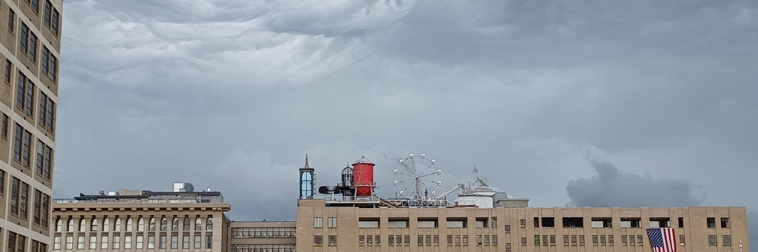
Near miss, a wedge tornado rolled through downtown west (St. Louis, MO) yesterday, moving just northwest of my building. Very close, but weirdly we didn't even get rain at our building, just a mile or two away.

May 17, 2025 at 2:42 PM
Near miss, a wedge tornado rolled through downtown west (St. Louis, MO) yesterday, moving just northwest of my building. Very close, but weirdly we didn't even get rain at our building, just a mile or two away.
Further attempts to stay positive and push the boundaries of X-ray imaging in plant biology, here's a maize root tip. Technical voxel resolution is 0.8um, field of view is about 1mm tall, using the 20X lens on my ZEISS Versa 520 XRM.
@danforthcenter.bsky.social
@zeiss-microscopy.bsky.social
@danforthcenter.bsky.social
@zeiss-microscopy.bsky.social
May 13, 2025 at 2:08 PM
Further attempts to stay positive and push the boundaries of X-ray imaging in plant biology, here's a maize root tip. Technical voxel resolution is 0.8um, field of view is about 1mm tall, using the 20X lens on my ZEISS Versa 520 XRM.
@danforthcenter.bsky.social
@zeiss-microscopy.bsky.social
@danforthcenter.bsky.social
@zeiss-microscopy.bsky.social
I'm constantly trying to get lost in my X-ray microscopy and forget the outside world. Here's a fly-through from an XRM scan of a fixed & contrast enhanced Beech tree bud, infested with nematodes.
@danforthcenter.bsky.social
@danforthcenter.bsky.social
April 3, 2025 at 10:44 PM
I'm constantly trying to get lost in my X-ray microscopy and forget the outside world. Here's a fly-through from an XRM scan of a fixed & contrast enhanced Beech tree bud, infested with nematodes.
@danforthcenter.bsky.social
@danforthcenter.bsky.social
In a continuing effort to focus on science, maize pollen fixed, contrast enhanced, and imaged with lab-based X-ray microscopy. Average diameter is ~90um. @danforthcenter.bsky.social
March 14, 2025 at 1:03 PM
In a continuing effort to focus on science, maize pollen fixed, contrast enhanced, and imaged with lab-based X-ray microscopy. Average diameter is ~90um. @danforthcenter.bsky.social
We all need our coping strategies, mine is Saturday afternoons at Stanley's.

February 15, 2025 at 9:58 PM
We all need our coping strategies, mine is Saturday afternoons at Stanley's.
I'm trying to stay positive and push imaging boundaries. The latest fascinating project is XRM imaging of Marchantia colonized by a mycorrhizal fungus. I'm sure there are false positives in this quick manual segmentation, but certainly some cool AMF structures as well. A satisfying first scan.
February 8, 2025 at 5:08 PM
I'm trying to stay positive and push imaging boundaries. The latest fascinating project is XRM imaging of Marchantia colonized by a mycorrhizal fungus. I'm sure there are false positives in this quick manual segmentation, but certainly some cool AMF structures as well. A satisfying first scan.
X-ray microscope imaging of AMF Rhizophagus irregularis colonized maize roots. Colonized roots were excavated and contrast enhanced with ePTA prior to XRM imaging at 1.3um pixel pitch. Still trying to get this to work in situ and avoid using osmium as a contrast agent.
February 1, 2025 at 5:31 PM
X-ray microscope imaging of AMF Rhizophagus irregularis colonized maize roots. Colonized roots were excavated and contrast enhanced with ePTA prior to XRM imaging at 1.3um pixel pitch. Still trying to get this to work in situ and avoid using osmium as a contrast agent.
A form most beautiful to me, a simple soybean flower visualized using X-ray microscopy.

January 26, 2025 at 5:40 PM
A form most beautiful to me, a simple soybean flower visualized using X-ray microscopy.
It's scary to be a scientist right now, the sheer magnitude of uncertainty can be paralyzing. I'm trying to find some level of calm, control what I can control.

January 25, 2025 at 9:45 PM
It's scary to be a scientist right now, the sheer magnitude of uncertainty can be paralyzing. I'm trying to find some level of calm, control what I can control.
Starting 2025 off with a beautiful XRM scan of a soybean root nodule, contrasted with ePTA. Lovely dataset for someone who's skilled at segmentation!
January 8, 2025 at 4:21 PM
Starting 2025 off with a beautiful XRM scan of a soybean root nodule, contrasted with ePTA. Lovely dataset for someone who's skilled at segmentation!
Another new collaboration, looking at developing tomato flowers using XRM. It will be exciting to see what the mutant looks like compared to this wild type.
December 18, 2024 at 3:29 PM
Another new collaboration, looking at developing tomato flowers using XRM. It will be exciting to see what the mutant looks like compared to this wild type.
Following up on my earlier post, XRM imaging of a collard green inflorescence. The real power of XRM imaging in plant biology is generating high resolution cell-level image data through the entire intact volume, in this case pollen-filled anthers surrounding the stigmatic surface.
December 14, 2024 at 5:01 PM
Following up on my earlier post, XRM imaging of a collard green inflorescence. The real power of XRM imaging in plant biology is generating high resolution cell-level image data through the entire intact volume, in this case pollen-filled anthers surrounding the stigmatic surface.
It's inflorescence Friday, so this is an XRM scan of young flower from a collard green, Susan Turner variety, collected by Antonio Brazelton. Fixed and contrast enhanced, technical voxel resolution of 1.4um. Imaging like this makes me happy, I need that right now.
December 13, 2024 at 3:47 PM
It's inflorescence Friday, so this is an XRM scan of young flower from a collard green, Susan Turner variety, collected by Antonio Brazelton. Fixed and contrast enhanced, technical voxel resolution of 1.4um. Imaging like this makes me happy, I need that right now.