


Captain Hooks
@captainhooks-lab.bsky.social
Motor circuits lab head @PittMed 🐭🧠🔬.
I would be remiss if I did not add that this project started long ago, with draft text and figures so clear in a folder optimistically named "PaperInAWeek20221019"
December 19, 2024 at 8:35 PM
I would be remiss if I did not add that this project started long ago, with draft text and figures so clear in a folder optimistically named "PaperInAWeek20221019"
While revising this paper, it was at bioRxiv and was cited in this nice review on bilateral sensorimotor function in striatum by Gómez-Ocádiz and Silberberg and that helped encourage us to finish the work: www.sciencedirect.com/science/arti... 24/24

Corticostriatal pathways for bilateral sensorimotor functions
Corticostriatal pathways are essential for a multitude of motor, sensory, cognitive, and affective functions. They are mediated by cortical pyramidal …
www.sciencedirect.com
December 19, 2024 at 8:35 PM
While revising this paper, it was at bioRxiv and was cited in this nice review on bilateral sensorimotor function in striatum by Gómez-Ocádiz and Silberberg and that helped encourage us to finish the work: www.sciencedirect.com/science/arti... 24/24
Frontal areas are then only split into sides because the constraints of development and anatomy divide the brain into hemispheres. 23/24
December 19, 2024 at 8:34 PM
Frontal areas are then only split into sides because the constraints of development and anatomy divide the brain into hemispheres. 23/24
in frontal areas than in sensory or motor ones b/c frontal cortex makes computations about an overall plan/decision for the whole animal. When deciding on what benefits the animal overall, it’s important to be of one mind and execute the plan in a coordinated manner. 22/24
December 19, 2024 at 8:33 PM
in frontal areas than in sensory or motor ones b/c frontal cortex makes computations about an overall plan/decision for the whole animal. When deciding on what benefits the animal overall, it’s important to be of one mind and execute the plan in a coordinated manner. 22/24
Do we get to speculate about why this is the case? Separate sensory and motor areas work fine when each is processing info appropriate to their own side of the body. But I conjecture that contralateral corticocortical and corticostriatal projections may play a larger role 21/24
December 19, 2024 at 8:33 PM
Do we get to speculate about why this is the case? Separate sensory and motor areas work fine when each is processing info appropriate to their own side of the body. But I conjecture that contralateral corticocortical and corticostriatal projections may play a larger role 21/24
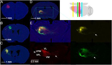
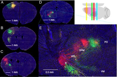
These were not as rich as the IT-type axons because corticothalamic projections only weakly cross the midline. But maybe we will be able to make some useful study of the differences of these two thalamic inputs in the future. 20/24
December 19, 2024 at 8:33 PM
These were not as rich as the IT-type axons because corticothalamic projections only weakly cross the midline. But maybe we will be able to make some useful study of the differences of these two thalamic inputs in the future. 20/24
We were also asked by the reviewers about the corticothalamic projections. Here we used some other mouse lines in L5B (PT-type projections, Sim1_KJ18_Cre) and L6 (CT-type projections, Ntsr1_GN220_Cre). 19/24
December 19, 2024 at 8:32 PM
We were also asked by the reviewers about the corticothalamic projections. Here we used some other mouse lines in L5B (PT-type projections, Sim1_KJ18_Cre) and L6 (CT-type projections, Ntsr1_GN220_Cre). 19/24
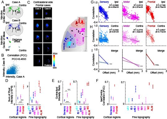
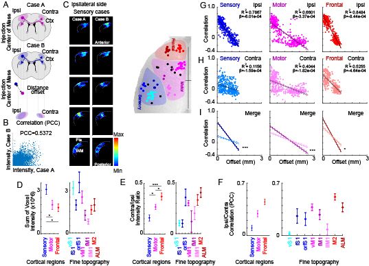
Again, we see that injections close to each other – low offset (in um) – are again pretty similar (7GHI, top). This is especially strong in frontal and sensory. But correlations across the midline were symmetrical/strong for frontal but not motor or sensory (7GHI, middle). 18/24
December 19, 2024 at 8:32 PM
Again, we see that injections close to each other – low offset (in um) – are again pretty similar (7GHI, top). This is especially strong in frontal and sensory. But correlations across the midline were symmetrical/strong for frontal but not motor or sensory (7GHI, middle). 18/24
The striatal comparison is similar to the result in cortex: frontal projections to the other side are stronger than motor which in turn is stronger than sensory (7E). 17/24
December 19, 2024 at 8:31 PM
The striatal comparison is similar to the result in cortex: frontal projections to the other side are stronger than motor which in turn is stronger than sensory (7E). 17/24
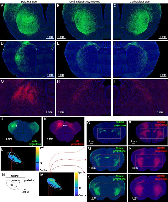
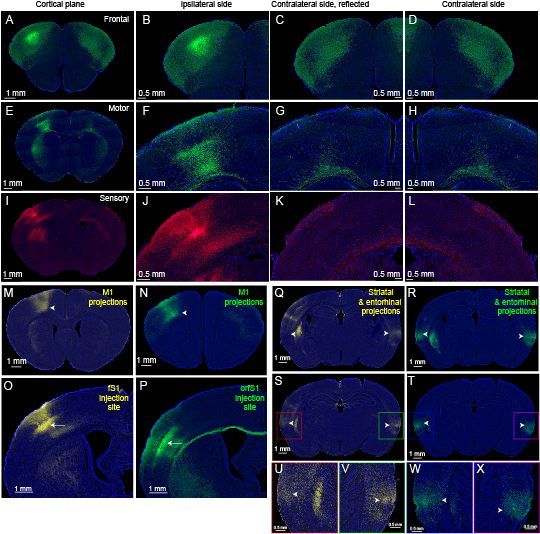
We made similar comparisons in striatum. These images of ipsi and contralateral frontal corticostriatal projections are quite pleasing to the eye. They are, as you might predict from cortex, the most symmetrical for more anterior projections. 16/24
December 19, 2024 at 8:31 PM
We made similar comparisons in striatum. These images of ipsi and contralateral frontal corticostriatal projections are quite pleasing to the eye. They are, as you might predict from cortex, the most symmetrical for more anterior projections. 16/24
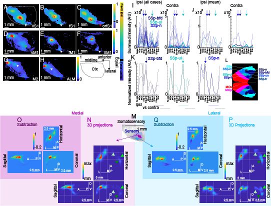
We even subdivided medial and lateral parts of S1 (suggested by a reviewer) and you can see how they differently project to the contralateral entorhinal areas (5MNOPQ). This figure was fun to make. 15/24
December 19, 2024 at 8:31 PM
We even subdivided medial and lateral parts of S1 (suggested by a reviewer) and you can see how they differently project to the contralateral entorhinal areas (5MNOPQ). This figure was fun to make. 15/24
So, we made 3D pictures of each brain, leaving only the detectable axons (suprathreshold voxels to make it sound more scholarly). We could average these together, subtract left from right, and show where projections are stronger or weaker on each side. This is nice (5A-H). 14/24
December 19, 2024 at 8:30 PM
So, we made 3D pictures of each brain, leaving only the detectable axons (suprathreshold voxels to make it sound more scholarly). We could average these together, subtract left from right, and show where projections are stronger or weaker on each side. This is nice (5A-H). 14/24
But the correlations across the midline followed a different pattern: these were strong for frontal but not motor or sensory (3GHI, middle). This finding (and the similar one in striatum) I think is pretty neat. 13/24
December 19, 2024 at 8:30 PM
But the correlations across the midline followed a different pattern: these were strong for frontal but not motor or sensory (3GHI, middle). This finding (and the similar one in striatum) I think is pretty neat. 13/24
This shows that injections close to each other – low offset (in um) – are pretty similar in the intensity of their correlation. They project to the same places ipsilaterally. This is especially strong in frontal and sensory. 12/24
December 19, 2024 at 8:30 PM
This shows that injections close to each other – low offset (in um) – are pretty similar in the intensity of their correlation. They project to the same places ipsilaterally. This is especially strong in frontal and sensory. 12/24
The cortical comparison shows that frontal projections to the other side are stronger than motor which in turn is stronger than sensory (3E). But even more than the strength, we were able to compare each injection to nearby injections from different mice (3GHI, top). 11/24
December 19, 2024 at 8:29 PM
The cortical comparison shows that frontal projections to the other side are stronger than motor which in turn is stronger than sensory (3E). But even more than the strength, we were able to compare each injection to nearby injections from different mice (3GHI, top). 11/24
Digital anatomy I think will let us play with the data in some fun ways. We can digitally make a mirror image to compare left/right brains in the same mouse. I suspect computational ability is beginning to outpace my ability to come up with what questions we should ask ... 10/24
December 19, 2024 at 8:29 PM
Digital anatomy I think will let us play with the data in some fun ways. We can digitally make a mirror image to compare left/right brains in the same mouse. I suspect computational ability is beginning to outpace my ability to come up with what questions we should ask ... 10/24
Alignment lets us define which voxels are in defined cortical and striatal areas, and using the same definition across mice, make quantitative, objective comparisons. 9/24
December 19, 2024 at 8:29 PM
Alignment lets us define which voxels are in defined cortical and striatal areas, and using the same definition across mice, make quantitative, objective comparisons. 9/24
Because we can do neat computational things like align the whole image stack to a standard brain, then we can compare injections across mice, tracking how close the injection sites were and also directly compare the axonal projections in the aligned brain space. 8/24
December 19, 2024 at 8:28 PM
Because we can do neat computational things like align the whole image stack to a standard brain, then we can compare injections across mice, tracking how close the injection sites were and also directly compare the axonal projections in the aligned brain space. 8/24
Focus on 3 parts of cortex: anterior mouse brain (frontal), motor, and somatosensory areas. I tried to stick to a red/purple/blue color scheme for these areas through the paper. Here we show where the injection was, with the ipsilateral and contralateral cortex in detail. 7/24
December 19, 2024 at 8:28 PM
Focus on 3 parts of cortex: anterior mouse brain (frontal), motor, and somatosensory areas. I tried to stick to a red/purple/blue color scheme for these areas through the paper. Here we show where the injection was, with the ipsilateral and contralateral cortex in detail. 7/24
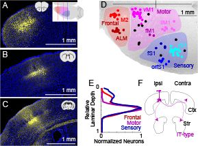
This line is pretty well restricted to layer 5 and we have a library of injections all over the dorsal cortex. As you might expect in anatomy work, we try to show some good examples of well labeled axons in a few examples. 6/24
December 19, 2024 at 8:28 PM
This line is pretty well restricted to layer 5 and we have a library of injections all over the dorsal cortex. As you might expect in anatomy work, we try to show some good examples of well labeled axons in a few examples. 6/24
We have a beautiful library of images from defined cell types, including the strong corticostriatal projection in layer 5 (we call these IT-type cells, we label them with a great transgenic mouse line, Tlx3_PL56_Cre). 5/24
December 19, 2024 at 8:27 PM
We have a beautiful library of images from defined cell types, including the strong corticostriatal projection in layer 5 (we call these IT-type cells, we label them with a great transgenic mouse line, Tlx3_PL56_Cre). 5/24
Striatum is generally involved in motivated movements, but many cortical areas, including primary sensory ones, send projections there. So how do these across the midline compare? Are they strong or weak? And do they project to comparable parts of the brain for each side? 4/24
December 19, 2024 at 8:27 PM
Striatum is generally involved in motivated movements, but many cortical areas, including primary sensory ones, send projections there. So how do these across the midline compare? Are they strong or weak? And do they project to comparable parts of the brain for each side? 4/24
But some projections cross the midline & connect to the other side. There's lots of anatomical examples in our data. So, we were curious about how cortical projections to contralateral cortex & basal ganglia, esp. striatum, compared across left and right sides of the body. 3/24
December 19, 2024 at 8:26 PM
But some projections cross the midline & connect to the other side. There's lots of anatomical examples in our data. So, we were curious about how cortical projections to contralateral cortex & basal ganglia, esp. striatum, compared across left and right sides of the body. 3/24
Generally, the human brain is organized in a criss-crossed manner with the left side of the brain seeing/touching the right side of the world & controling movement of the body's right side. This is fascinating when we first learn it in school, but not well understood why. 2/24
December 19, 2024 at 8:26 PM
Generally, the human brain is organized in a criss-crossed manner with the left side of the brain seeing/touching the right side of the world & controling movement of the body's right side. This is fascinating when we first learn it in school, but not well understood why. 2/24