


Andrea Vettiger
@avettiger.bsky.social
Assistant Professor
@dmf-unil.bsky.social
https://wp.unil.ch/vettiger-lab/
Postdoc Bernhardt lab | PhD Basler lab.
Shining light on small bugs to reveal their amazing biology! #elongasome #divisome #T6SS
@dmf-unil.bsky.social
https://wp.unil.ch/vettiger-lab/
Postdoc Bernhardt lab | PhD Basler lab.
Shining light on small bugs to reveal their amazing biology! #elongasome #divisome #T6SS
Congrats, that’s great!
April 4, 2025 at 1:47 PM
Congrats, that’s great!
This work was carried out largely as part of a fantastic postdoc in Tom Bernhardt's lab at @harvardmed.bsky.social. @navarropaula.bsky.social and myself both started our own labs at @dmf-unil.bsky.social last year and just received some funding. Job post on related topics will follow soon 😃 (12/12)
April 3, 2025 at 10:13 PM
This work was carried out largely as part of a fantastic postdoc in Tom Bernhardt's lab at @harvardmed.bsky.social. @navarropaula.bsky.social and myself both started our own labs at @dmf-unil.bsky.social last year and just received some funding. Job post on related topics will follow soon 😃 (12/12)
Based on AlphaFold prediction, this PBP1b-FtsA interaction is conserved among many Enterobactericae (in red) but not beyond. However, similar intrinsically disordered domains are found on most PBP1b and may offer a way to regulate aPBP activity independent of Lpo's. (11/12)

April 3, 2025 at 10:13 PM
Based on AlphaFold prediction, this PBP1b-FtsA interaction is conserved among many Enterobactericae (in red) but not beyond. However, similar intrinsically disordered domains are found on most PBP1b and may offer a way to regulate aPBP activity independent of Lpo's. (11/12)
In the previous movie FtsZ (yellow) gets displaced from FtsA (unlabeled) upon addition of the wt N-terminal peptide of PBP1b (magenta), but not the R6E point mutant 👇 (10/12)
April 3, 2025 at 10:13 PM
In the previous movie FtsZ (yellow) gets displaced from FtsA (unlabeled) upon addition of the wt N-terminal peptide of PBP1b (magenta), but not the R6E point mutant 👇 (10/12)
To our surprise the peptide of PBP1b was predicted to interact with the same residue on FtsA than FtsZ. This may suggest a competitive binding for FtsZ and PBP1b to FtsA. Thanks to Roman Hajdu in @nartimsoole.bsky.social lab, we find evidence for this in supported lipid bilayer assays 🧪🔬 (9/12)
April 3, 2025 at 10:13 PM
To our surprise the peptide of PBP1b was predicted to interact with the same residue on FtsA than FtsZ. This may suggest a competitive binding for FtsZ and PBP1b to FtsA. Thanks to Roman Hajdu in @nartimsoole.bsky.social lab, we find evidence for this in supported lipid bilayer assays 🧪🔬 (9/12)
But how does the N-terminal peptide of PBP1b mediate septal recruitment? AlphaFold screen performed by @ernstschmid.bsky.social in Johannes Walker's lab predicted an interaction with FtsA via a salt bridge between FtsA(E303) and PBP1b(R6) 🖥️ (8/12)

April 3, 2025 at 10:13 PM
But how does the N-terminal peptide of PBP1b mediate septal recruitment? AlphaFold screen performed by @ernstschmid.bsky.social in Johannes Walker's lab predicted an interaction with FtsA via a salt bridge between FtsA(E303) and PBP1b(R6) 🖥️ (8/12)
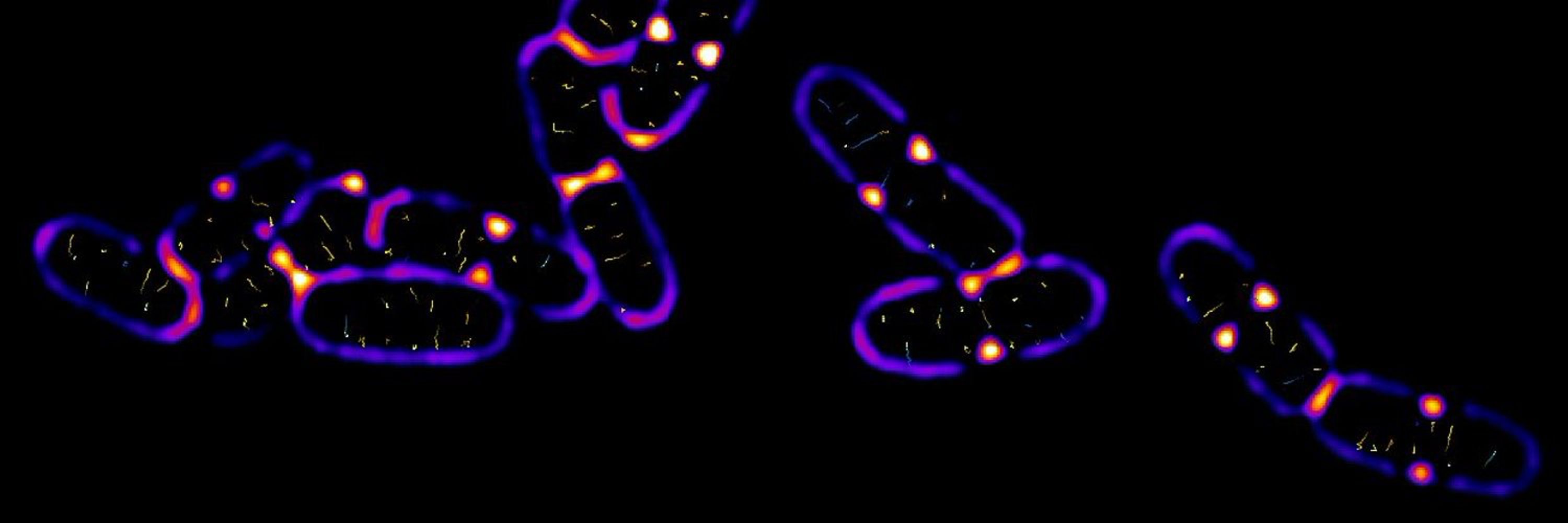
The GFP fusion to the PBP1b-alpha isoform indeed does display a biased septal localization pattern unlike the shorter gamma isoform. This is supported by previous findings using immunofluorescence by Tanneke den Blaauwen and Waldemar Vollmer's group (onlinelibrary.wiley.com/doi/10.1111/...) ✅ (7/12)
https://onlinelibrary.wiley.com/doi/10.1111/j.1365-2958.2006.05280.x✅
April 3, 2025 at 10:13 PM
The GFP fusion to the PBP1b-alpha isoform indeed does display a biased septal localization pattern unlike the shorter gamma isoform. This is supported by previous findings using immunofluorescence by Tanneke den Blaauwen and Waldemar Vollmer's group (onlinelibrary.wiley.com/doi/10.1111/...) ✅ (7/12)
Interestingly, septal fortification of PBP1b doesn't seem to depend on its canonical Lpo activator, LpoB. However, it is dependent on the N-terminal cytosolic peptide of PBP1b, which was omitted in previous GFP fusions resulting in uniform IM labeling pattern 🤔 (6/12)

April 3, 2025 at 10:13 PM
Interestingly, septal fortification of PBP1b doesn't seem to depend on its canonical Lpo activator, LpoB. However, it is dependent on the N-terminal cytosolic peptide of PBP1b, which was omitted in previous GFP fusions resulting in uniform IM labeling pattern 🤔 (6/12)
This lysis phenotype is specific for cells undergoing division. When inhibiting divisome formation with SulA expression, cells lacking PBP1b lyse significantly less, and when they do, they display smaller lesions which are likely attributed to the impaired side wall repair 🛠️ (6/12)
April 3, 2025 at 10:13 PM
This lysis phenotype is specific for cells undergoing division. When inhibiting divisome formation with SulA expression, cells lacking PBP1b lyse significantly less, and when they do, they display smaller lesions which are likely attributed to the impaired side wall repair 🛠️ (6/12)
Having such compromised septa leads to catastrophic lysis events under osmotically stressed conditions originating from the division site (green = released DNA) 🌊🔬(5/12)
April 3, 2025 at 10:13 PM
Having such compromised septa leads to catastrophic lysis events under osmotically stressed conditions originating from the division site (green = released DNA) 🌊🔬(5/12)
And their septal cell walls show compromised integrity (more pores and less stiff) when measured by AFM ⚛️🔬 (4/12).

April 3, 2025 at 10:13 PM
And their septal cell walls show compromised integrity (more pores and less stiff) when measured by AFM ⚛️🔬 (4/12).
Using in situ cryo-ET (as always performed by @navarropaula.bsky.social) we show that cells lacking PBP1b fail to produce a so-called septal PG wedge (highlighted by the red arrow) ❄️🔬 (3/12)

April 3, 2025 at 10:13 PM
Using in situ cryo-ET (as always performed by @navarropaula.bsky.social) we show that cells lacking PBP1b fail to produce a so-called septal PG wedge (highlighted by the red arrow) ❄️🔬 (3/12)
aPBPs are a major cell wall synthase family and have been implemented in PG repair and fortification. Additionally, PBP1b has been proposed to play a crucial role in cell division however its precise role has remained obscure. (2/12)

April 3, 2025 at 10:13 PM
aPBPs are a major cell wall synthase family and have been implemented in PG repair and fortification. Additionally, PBP1b has been proposed to play a crucial role in cell division however its precise role has remained obscure. (2/12)