


Elio Abbondanzieri
@abbondanzieri.bsky.social
Dad, Husband, Biophysicist, Tinkerer
"It's 106 miles to the Vatican, we got a full tank of gas, half a pack of incense, it's dark and we're wearing sunglasses. Hit it!"

November 13, 2025 at 4:49 PM
"It's 106 miles to the Vatican, we got a full tank of gas, half a pack of incense, it's dark and we're wearing sunglasses. Hit it!"
It is an important reminder that cells aren't just bags of chemicals reacting with each other, they are complex tiny physical machines with intricate internal architectures that dictate what they do. Thanks for reading this far!

October 22, 2025 at 7:21 PM
It is an important reminder that cells aren't just bags of chemicals reacting with each other, they are complex tiny physical machines with intricate internal architectures that dictate what they do. Thanks for reading this far!
So taking our data all together, we see that to understand what H-NS is doing, we can't just ask if it is present and if it is bound to the DNA. We have to go deeper look at the structure the H-NS creates in the genome.

October 22, 2025 at 7:21 PM
So taking our data all together, we see that to understand what H-NS is doing, we can't just ask if it is present and if it is bound to the DNA. We have to go deeper look at the structure the H-NS creates in the genome.
When we asked what parts of the genome are being downregulated or upregulated, we see that downregulated genes correlated with H-NS binding and the Hi-C peaks. By contrast, upregulated genes are weakly anti-correlated with these things.

October 22, 2025 at 7:21 PM
When we asked what parts of the genome are being downregulated or upregulated, we see that downregulated genes correlated with H-NS binding and the Hi-C peaks. By contrast, upregulated genes are weakly anti-correlated with these things.
We also performed RNAseq, a technique that lets us monitor gene expression across the cell. The ability of H-NS to repress gene expression was much stronger during stationary phase. Surprisingly, H-NS was able to broadly upregulate numerous genes during stationary phase.

October 22, 2025 at 7:21 PM
We also performed RNAseq, a technique that lets us monitor gene expression across the cell. The ability of H-NS to repress gene expression was much stronger during stationary phase. Surprisingly, H-NS was able to broadly upregulate numerous genes during stationary phase.
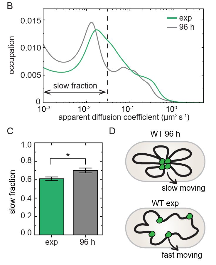
The Biteen lab also showed H-NS was excluding large protein assemblies from accessing the middle of the cell during stationary phase. So H-NS was forming a stable structure in the cell that affected how objects diffuse around the cytoplasm.

October 22, 2025 at 7:21 PM
The Biteen lab also showed H-NS was excluding large protein assemblies from accessing the middle of the cell during stationary phase. So H-NS was forming a stable structure in the cell that affected how objects diffuse around the cytoplasm.
We wanted to know what this meant, and we turned to Lauren McCarthy from the @juliebiteenlab.bsky.social lab to do single molecule analysis inside of the cells. Lauren McCarthy was able to demonstrate with other students that H-NS was anchored down more strongly in stationary phase.
October 22, 2025 at 7:21 PM
We wanted to know what this meant, and we turned to Lauren McCarthy from the @juliebiteenlab.bsky.social lab to do single molecule analysis inside of the cells. Lauren McCarthy was able to demonstrate with other students that H-NS was anchored down more strongly in stationary phase.
And the parts of the genome that were packed into this core? We can identify that by integrating the peaks in the Hi-C contact maps. It turned out to be almost identical where on the genome H-NS was bound detected by ChIP-seq.

October 22, 2025 at 7:21 PM
And the parts of the genome that were packed into this core? We can identify that by integrating the peaks in the Hi-C contact maps. It turned out to be almost identical where on the genome H-NS was bound detected by ChIP-seq.
We saw a characteristic pattern that was consistent with a few disparate regions of the DNA around the genome were "pinched" together during stationary phase into a dense core. This dense core only formed if H-NS was present and the cell was in stationary phase.

October 22, 2025 at 7:21 PM
We saw a characteristic pattern that was consistent with a few disparate regions of the DNA around the genome were "pinched" together during stationary phase into a dense core. This dense core only formed if H-NS was present and the cell was in stationary phase.
Importantly, she was able to adapt the technique to work in stationary phase, which has been a barrier for most labs. The raw data was challenging to visualize at first, but we were able to use algorithms designed for image analysis to isolate the long-range interactions.

October 22, 2025 at 7:21 PM
Importantly, she was able to adapt the technique to work in stationary phase, which has been a barrier for most labs. The raw data was challenging to visualize at first, but we were able to use algorithms designed for image analysis to isolate the long-range interactions.
Here we were lucky to be collaborating with Xindan Wang, a pioneer in using high-throughput sequencing techniques. Lindsey Way, a graduate student in the Wang Lab, was able to use Hi-C to generate a heat map of all the contacts between different DNA regions.

October 22, 2025 at 7:21 PM
Here we were lucky to be collaborating with Xindan Wang, a pioneer in using high-throughput sequencing techniques. Lindsey Way, a graduate student in the Wang Lab, was able to use Hi-C to generate a heat map of all the contacts between different DNA regions.
What's more, when we look at where H-NS binds to DNA using ChIP-seq, we get a very similar picture in both stationary phase and the fast growing cells (i.e. exponential phase). So why would it be better at turning genes off in stationary phase?

October 22, 2025 at 7:21 PM
What's more, when we look at where H-NS binds to DNA using ChIP-seq, we get a very similar picture in both stationary phase and the fast growing cells (i.e. exponential phase). So why would it be better at turning genes off in stationary phase?
Excited to share a new preprint from a team of researchers I am part of (funded by an NIH grant). The paper helps us understand a question I have been interested in for a while: why are bacteria so resilient?
www.biorxiv.org/content/10.1...
@meyerroc.bsky.social
@juliebiteenlab.bsky.social
www.biorxiv.org/content/10.1...
@meyerroc.bsky.social
@juliebiteenlab.bsky.social
October 22, 2025 at 7:21 PM
Excited to share a new preprint from a team of researchers I am part of (funded by an NIH grant). The paper helps us understand a question I have been interested in for a while: why are bacteria so resilient?
www.biorxiv.org/content/10.1...
@meyerroc.bsky.social
@juliebiteenlab.bsky.social
www.biorxiv.org/content/10.1...
@meyerroc.bsky.social
@juliebiteenlab.bsky.social
Not sure what's going on with the Atlanta FED's GdpNOW model, but it has been predicting a large contraction for months. Then at some point today, the old numbers were replaced by a single point claiming the model predicts the economy is growing at a high rate.
www.atlantafed.org/cqer/researc...
www.atlantafed.org/cqer/researc...


April 30, 2025 at 5:17 PM
Not sure what's going on with the Atlanta FED's GdpNOW model, but it has been predicting a large contraction for months. Then at some point today, the old numbers were replaced by a single point claiming the model predicts the economy is growing at a high rate.
www.atlantafed.org/cqer/researc...
www.atlantafed.org/cqer/researc...
Atlanta FED's GDPNow model is predicting the US GPD already started shrinking in March, before tariffs were announced. Models like this aren't perfect but they aren't susceptible to herding like human forecasters are.
www.atlantafed.org/cqer/researc...
www.atlantafed.org/cqer/researc...

April 9, 2025 at 4:12 PM
Atlanta FED's GDPNow model is predicting the US GPD already started shrinking in March, before tariffs were announced. Models like this aren't perfect but they aren't susceptible to herding like human forecasters are.
www.atlantafed.org/cqer/researc...
www.atlantafed.org/cqer/researc...
The cold weather couldn't keep people away from today's Stand up for Science rally at RIT.
@standupforscience.bsky.social
@standupforscience.bsky.social


March 7, 2025 at 6:25 PM
The cold weather couldn't keep people away from today's Stand up for Science rally at RIT.
@standupforscience.bsky.social
@standupforscience.bsky.social
It's nice to finally have a movie that brings to life that classic EM image of a lysed E. coli.

March 3, 2025 at 10:41 AM
It's nice to finally have a movie that brings to life that classic EM image of a lysed E. coli.
So we used the same technique to compare bacteria with and without a silica coating. We see bacteria with the coating are trapped less stiffly than bacteria without a coating, presumably because the scattering force on the beads goes up faster than the gradient force.


December 11, 2024 at 6:49 PM
So we used the same technique to compare bacteria with and without a silica coating. We see bacteria with the coating are trapped less stiffly than bacteria without a coating, presumably because the scattering force on the beads goes up faster than the gradient force.
Oh, and one bonus story: We also checked for the silica coating using optical tweezers from @lumicks.bsky.social. We trapped beads of similar sizes made of different materials to show we could detect changes in the refractive index by looking at changes in the stiffness as a function of diameter...

December 11, 2024 at 6:43 PM
Oh, and one bonus story: We also checked for the silica coating using optical tweezers from @lumicks.bsky.social. We trapped beads of similar sizes made of different materials to show we could detect changes in the refractive index by looking at changes in the stiffness as a function of diameter...
But even more exciting is the fact that these glass-coated cells are apparently still alive. Lynn used an alamarBlue assay to see when the cells stopped being metabolically active and they kept going for months after encapsulation. So the cells could potentially express genes after encapsulation.

December 11, 2024 at 4:36 PM
But even more exciting is the fact that these glass-coated cells are apparently still alive. Lynn used an alamarBlue assay to see when the cells stopped being metabolically active and they kept going for months after encapsulation. So the cells could potentially express genes after encapsulation.
E. coli have interesting properties that could be useful for developing photonic devices. They are rod-shaped instead of round, so how they scatter light depends on their alignment. We think there may be some immediate applications for microlenses with this unusual shape.


December 11, 2024 at 4:26 PM
E. coli have interesting properties that could be useful for developing photonic devices. They are rod-shaped instead of round, so how they scatter light depends on their alignment. We think there may be some immediate applications for microlenses with this unusual shape.
Not only did the scattering pattern match the theory, but the silica coated bacteria scattered a *lot* more light than the uncoated cells. We had transformed bacteria into tiny microlenses!


December 11, 2024 at 4:19 PM
Not only did the scattering pattern match the theory, but the silica coated bacteria scattered a *lot* more light than the uncoated cells. We had transformed bacteria into tiny microlenses!
Could we measure this scattering on a microscope? Yes! We modified a microscope designed for TIRF illumination so that we could shine light at very shallow angles onto the cells. We used fluorescent dyes to see the jets of light coming out of the cells, and they matched the theory beautifully!

December 11, 2024 at 4:06 PM
Could we measure this scattering on a microscope? Yes! We modified a microscope designed for TIRF illumination so that we could shine light at very shallow angles onto the cells. We used fluorescent dyes to see the jets of light coming out of the cells, and they matched the theory beautifully!
That student, Lynn Sydor, then wanted to see if this glass coating would change the optical properties of the cells. The coating was very thin (~10 nm), so you might assume any changes would be minor. However, modeling suggested this thin layer could make a large difference in how light scattered.

December 11, 2024 at 4:00 PM
That student, Lynn Sydor, then wanted to see if this glass coating would change the optical properties of the cells. The coating was very thin (~10 nm), so you might assume any changes would be minor. However, modeling suggested this thin layer could make a large difference in how light scattered.
A tenacious and clever graduate student in Anne's lab then set our to see if these enzymes could produce glass on E. coli cells. This was very difficult to measure, and she ended up using a variety of techniques (X-Ray Powder Diffraction, immunofluorescence, and electron microscopy) to show they do!

December 11, 2024 at 3:54 PM
A tenacious and clever graduate student in Anne's lab then set our to see if these enzymes could produce glass on E. coli cells. This was very difficult to measure, and she ended up using a variety of techniques (X-Ray Powder Diffraction, immunofluorescence, and electron microscopy) to show they do!