


The X-Vet
@xvetradiology.bsky.social
X-rays, ultrasounds, CT scans, and MRIs from a variety of animals.
Discussion of animal anatomy, medical cases and imaging techniques.
Perfect for veterinarians, students, and animal enthusiasts who are curious about the imaging side of animal care.
Discussion of animal anatomy, medical cases and imaging techniques.
Perfect for veterinarians, students, and animal enthusiasts who are curious about the imaging side of animal care.
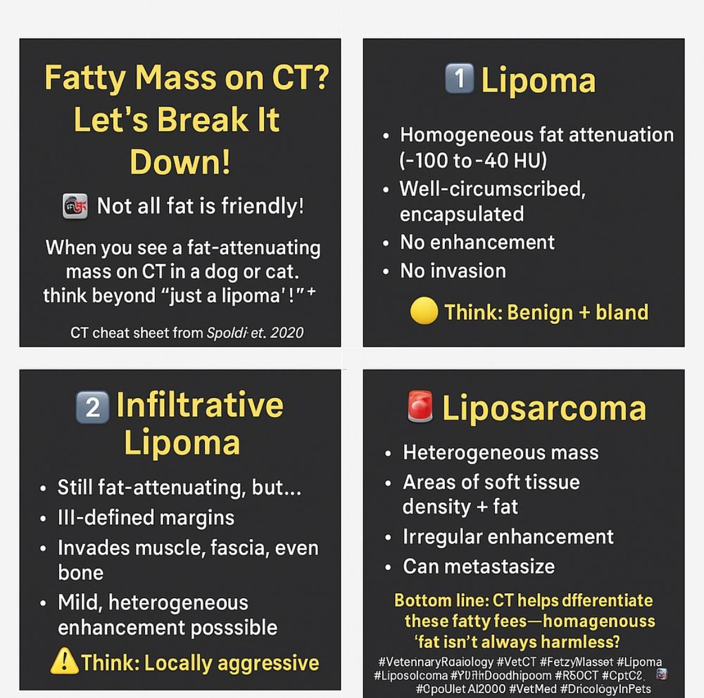
Fatty Mass on CT? Let’s Break It Down!
Not all fat is friendly! When you see a fat-attenuating mass on CT in a dog or cat, think beyond “just a lipoma”!
Here’s your CT cheat sheet from Spoldi et al., 2020
Bottom line: CT helps differentiate these fatty foes—homogeneous fat isn’t always harmless!#vet
Not all fat is friendly! When you see a fat-attenuating mass on CT in a dog or cat, think beyond “just a lipoma”!
Here’s your CT cheat sheet from Spoldi et al., 2020
Bottom line: CT helps differentiate these fatty foes—homogeneous fat isn’t always harmless!#vet

May 2, 2025 at 8:16 AM
Fatty Mass on CT? Let’s Break It Down!
Not all fat is friendly! When you see a fat-attenuating mass on CT in a dog or cat, think beyond “just a lipoma”!
Here’s your CT cheat sheet from Spoldi et al., 2020
Bottom line: CT helps differentiate these fatty foes—homogeneous fat isn’t always harmless!#vet
Not all fat is friendly! When you see a fat-attenuating mass on CT in a dog or cat, think beyond “just a lipoma”!
Here’s your CT cheat sheet from Spoldi et al., 2020
Bottom line: CT helps differentiate these fatty foes—homogeneous fat isn’t always harmless!#vet
Today is World Veterinary Day!
As a vet at The X-Vet, I feel so proud to be part of this amazing profession.
Veterinary medicine is about science, yes, but it’s also about compassion, patience, and a whole lot of love.
www.instagram.com/the_x_vet?ig...
As a vet at The X-Vet, I feel so proud to be part of this amazing profession.
Veterinary medicine is about science, yes, but it’s also about compassion, patience, and a whole lot of love.
www.instagram.com/the_x_vet?ig...

April 26, 2025 at 3:06 PM
Today is World Veterinary Day!
As a vet at The X-Vet, I feel so proud to be part of this amazing profession.
Veterinary medicine is about science, yes, but it’s also about compassion, patience, and a whole lot of love.
www.instagram.com/the_x_vet?ig...
As a vet at The X-Vet, I feel so proud to be part of this amazing profession.
Veterinary medicine is about science, yes, but it’s also about compassion, patience, and a whole lot of love.
www.instagram.com/the_x_vet?ig...
How to Spot the Gallbladder on MRI
You’re looking for a bright (✨ hyperintense) oval/pear-shaped thing between the liver lobes — yep, that’s the gallbladder! It’s full of bile, so it lights up like a pro on T2.
• Normal gallbladders can be bilobed — it’s cool, it’s normal!!
#vetimaging #vetmri
You’re looking for a bright (✨ hyperintense) oval/pear-shaped thing between the liver lobes — yep, that’s the gallbladder! It’s full of bile, so it lights up like a pro on T2.
• Normal gallbladders can be bilobed — it’s cool, it’s normal!!
#vetimaging #vetmri

April 8, 2025 at 6:30 PM
How to Spot the Gallbladder on MRI
You’re looking for a bright (✨ hyperintense) oval/pear-shaped thing between the liver lobes — yep, that’s the gallbladder! It’s full of bile, so it lights up like a pro on T2.
• Normal gallbladders can be bilobed — it’s cool, it’s normal!!
#vetimaging #vetmri
You’re looking for a bright (✨ hyperintense) oval/pear-shaped thing between the liver lobes — yep, that’s the gallbladder! It’s full of bile, so it lights up like a pro on T2.
• Normal gallbladders can be bilobed — it’s cool, it’s normal!!
#vetimaging #vetmri
Perinephric cysts are fluid-filled sacs that develop around the kidneys, often incidental findings
🔹 Anechoic fluid-filled structures surrounding the kidney
🔹 Well-defined, thin-walled, and non-vascular on Doppler
🔹 May cause kidney displacement or compression if large #vetultrasound #vetimaging
🔹 Anechoic fluid-filled structures surrounding the kidney
🔹 Well-defined, thin-walled, and non-vascular on Doppler
🔹 May cause kidney displacement or compression if large #vetultrasound #vetimaging


March 30, 2025 at 9:14 PM
Perinephric cysts are fluid-filled sacs that develop around the kidneys, often incidental findings
🔹 Anechoic fluid-filled structures surrounding the kidney
🔹 Well-defined, thin-walled, and non-vascular on Doppler
🔹 May cause kidney displacement or compression if large #vetultrasound #vetimaging
🔹 Anechoic fluid-filled structures surrounding the kidney
🔹 Well-defined, thin-walled, and non-vascular on Doppler
🔹 May cause kidney displacement or compression if large #vetultrasound #vetimaging
The coeliac artery (*) arises from the aorta caudal to the diaphragm and gives rise to the hepatic, splenic and left gastric arteries. The cranial mesenteric artery (#) emerges slightly caudal, providing vascular supply to the small intestine and part of the large intestine. #vetanatomy #vetmri

March 29, 2025 at 2:54 PM
The coeliac artery (*) arises from the aorta caudal to the diaphragm and gives rise to the hepatic, splenic and left gastric arteries. The cranial mesenteric artery (#) emerges slightly caudal, providing vascular supply to the small intestine and part of the large intestine. #vetanatomy #vetmri
Radiographs of cats with pulmonary tuberculosis typically reveal diffuse or nodular lung patterns, often resembling bronchopneumonia. Lesions may appear as interstitial or alveolar infiltrates.
Enlarged tracheobronchial lymph nodes and pleural effusion can also be present.
#vet #vetimaging #vetrad
Enlarged tracheobronchial lymph nodes and pleural effusion can also be present.
#vet #vetimaging #vetrad


February 27, 2025 at 10:41 PM
Radiographs of cats with pulmonary tuberculosis typically reveal diffuse or nodular lung patterns, often resembling bronchopneumonia. Lesions may appear as interstitial or alveolar infiltrates.
Enlarged tracheobronchial lymph nodes and pleural effusion can also be present.
#vet #vetimaging #vetrad
Enlarged tracheobronchial lymph nodes and pleural effusion can also be present.
#vet #vetimaging #vetrad
Left atrial mass presumptive myxoma
Myxomas are rare, benign primary cardiac tumors in dogs, most commonly found in the left atrium. Despite their benign nature, they can cause significant hemodynamic effects by obstructing blood flow at the mitral valve, leading to signs of congestive heart failure
Myxomas are rare, benign primary cardiac tumors in dogs, most commonly found in the left atrium. Despite their benign nature, they can cause significant hemodynamic effects by obstructing blood flow at the mitral valve, leading to signs of congestive heart failure


February 17, 2025 at 9:36 PM
Left atrial mass presumptive myxoma
Myxomas are rare, benign primary cardiac tumors in dogs, most commonly found in the left atrium. Despite their benign nature, they can cause significant hemodynamic effects by obstructing blood flow at the mitral valve, leading to signs of congestive heart failure
Myxomas are rare, benign primary cardiac tumors in dogs, most commonly found in the left atrium. Despite their benign nature, they can cause significant hemodynamic effects by obstructing blood flow at the mitral valve, leading to signs of congestive heart failure
Ectopic thyroid carcinoma should be considered in the differential diagnosis when a mass in the basihyoid region is present.
They were seen as oval masses centered on the basihyoid bone with associated bone lysis.
onlinelibrary.wiley.com/doi/10.1111/...
#vet #vetct #xvet #vetimaging #dog
They were seen as oval masses centered on the basihyoid bone with associated bone lysis.
onlinelibrary.wiley.com/doi/10.1111/...
#vet #vetct #xvet #vetimaging #dog


February 14, 2025 at 11:54 AM
Ectopic thyroid carcinoma should be considered in the differential diagnosis when a mass in the basihyoid region is present.
They were seen as oval masses centered on the basihyoid bone with associated bone lysis.
onlinelibrary.wiley.com/doi/10.1111/...
#vet #vetct #xvet #vetimaging #dog
They were seen as oval masses centered on the basihyoid bone with associated bone lysis.
onlinelibrary.wiley.com/doi/10.1111/...
#vet #vetct #xvet #vetimaging #dog
The muscularis layer of the gastrointestinal tract appears as a hypoechoic (dark) layer between the more echogenic submucosa and serosa.
Thickening of this layer can indicate inflammatory bowel disease, lymphoma, or other infiltrative diseases, making its assessment important.
#vet #vetimaging #xvet
Thickening of this layer can indicate inflammatory bowel disease, lymphoma, or other infiltrative diseases, making its assessment important.
#vet #vetimaging #xvet

February 8, 2025 at 3:07 PM
The muscularis layer of the gastrointestinal tract appears as a hypoechoic (dark) layer between the more echogenic submucosa and serosa.
Thickening of this layer can indicate inflammatory bowel disease, lymphoma, or other infiltrative diseases, making its assessment important.
#vet #vetimaging #xvet
Thickening of this layer can indicate inflammatory bowel disease, lymphoma, or other infiltrative diseases, making its assessment important.
#vet #vetimaging #xvet
For a CT urography protocol in dogs, the excretory phase is typically acquired at 3 minutes post-contrast injection, though some protocols extend it to 5–10 minutes for better visualization of ureters, especially in cases of delayed excretion.


January 30, 2025 at 9:56 PM
For a CT urography protocol in dogs, the excretory phase is typically acquired at 3 minutes post-contrast injection, though some protocols extend it to 5–10 minutes for better visualization of ureters, especially in cases of delayed excretion.
Answer: Renal carcinoma
Renal carcinomas typically appear as large, irregularly shaped masses with heterogeneous contrast enhancement due to areas of necrosis or hemorrhage.
#vet #vetimaging #vetradiology #vetct #xvet #renalcarcinoma #dogct
Renal carcinomas typically appear as large, irregularly shaped masses with heterogeneous contrast enhancement due to areas of necrosis or hemorrhage.
#vet #vetimaging #vetradiology #vetct #xvet #renalcarcinoma #dogct

January 28, 2025 at 9:29 PM
Answer: Renal carcinoma
Renal carcinomas typically appear as large, irregularly shaped masses with heterogeneous contrast enhancement due to areas of necrosis or hemorrhage.
#vet #vetimaging #vetradiology #vetct #xvet #renalcarcinoma #dogct
Renal carcinomas typically appear as large, irregularly shaped masses with heterogeneous contrast enhancement due to areas of necrosis or hemorrhage.
#vet #vetimaging #vetradiology #vetct #xvet #renalcarcinoma #dogct
In dogs, discospondylitis appears on MRI as hyperintense on T2 and STIR, highlighting inflammation and edema in the affected intervertebral disc, adjacent vertebral endplates, and surrounding soft tissues. T1 shows hypointensity with post-contrast enhancement revealing marked inflammation.
#vet
#vet


January 16, 2025 at 8:25 PM
In dogs, discospondylitis appears on MRI as hyperintense on T2 and STIR, highlighting inflammation and edema in the affected intervertebral disc, adjacent vertebral endplates, and surrounding soft tissues. T1 shows hypointensity with post-contrast enhancement revealing marked inflammation.
#vet
#vet
The medial pterygoid muscle is one of the primary muscles of mastication, and It works in conjunction with other masticatory muscles to aid in the closing of the jaw and side-to-side jaw movements, essential for chewing.
#vetradiology #xvet #vetmri #vetspecialists #vetanatomy #dog #vetimaging
#vetradiology #xvet #vetmri #vetspecialists #vetanatomy #dog #vetimaging

January 5, 2025 at 11:34 PM
The medial pterygoid muscle is one of the primary muscles of mastication, and It works in conjunction with other masticatory muscles to aid in the closing of the jaw and side-to-side jaw movements, essential for chewing.
#vetradiology #xvet #vetmri #vetspecialists #vetanatomy #dog #vetimaging
#vetradiology #xvet #vetmri #vetspecialists #vetanatomy #dog #vetimaging
Pericardial effusion
Radiographs are a valuable diagnostic tool for identifying pericardial effusion in dogs.
This condition, characterized by fluid accumulation within the pericardial sac, often appears on X-rays as an enlarged, globoid-shaped cardiac silhouette with sharply defined borders.
Radiographs are a valuable diagnostic tool for identifying pericardial effusion in dogs.
This condition, characterized by fluid accumulation within the pericardial sac, often appears on X-rays as an enlarged, globoid-shaped cardiac silhouette with sharply defined borders.


January 1, 2025 at 11:10 PM
Pericardial effusion
Radiographs are a valuable diagnostic tool for identifying pericardial effusion in dogs.
This condition, characterized by fluid accumulation within the pericardial sac, often appears on X-rays as an enlarged, globoid-shaped cardiac silhouette with sharply defined borders.
Radiographs are a valuable diagnostic tool for identifying pericardial effusion in dogs.
This condition, characterized by fluid accumulation within the pericardial sac, often appears on X-rays as an enlarged, globoid-shaped cardiac silhouette with sharply defined borders.
Answer: VD view is best to assess the accessory lung lobe
Check this paper out for further info!
Check this paper out for further info!


December 11, 2024 at 8:24 PM
Answer: VD view is best to assess the accessory lung lobe
Check this paper out for further info!
Check this paper out for further info!
Answer: the most common fidning in dogs with SRMA is paravertebral musculature changes
The most common MRI findings were paravertebral muscle changes (30/53; 56.6%), meningeal contrast enhancement (13/41; 31.7%) and spinal cord parenchymal T2-W hyperintensity (15/53; 28.3%).
#vet #vetimaging #mri
The most common MRI findings were paravertebral muscle changes (30/53; 56.6%), meningeal contrast enhancement (13/41; 31.7%) and spinal cord parenchymal T2-W hyperintensity (15/53; 28.3%).
#vet #vetimaging #mri


December 8, 2024 at 10:39 PM
Answer: the most common fidning in dogs with SRMA is paravertebral musculature changes
The most common MRI findings were paravertebral muscle changes (30/53; 56.6%), meningeal contrast enhancement (13/41; 31.7%) and spinal cord parenchymal T2-W hyperintensity (15/53; 28.3%).
#vet #vetimaging #mri
The most common MRI findings were paravertebral muscle changes (30/53; 56.6%), meningeal contrast enhancement (13/41; 31.7%) and spinal cord parenchymal T2-W hyperintensity (15/53; 28.3%).
#vet #vetimaging #mri
Laryngeal chondrosarcoma in dogs is a rare malignant tumor originating from the laryngeal cartilage.
Radiographs may reveal soft tissue swelling or mineralization within the larynx, but advanced imaging is often required for more detailed assessment.
#vetradiology #xvetradiology #larynx #vet #xrays
Radiographs may reveal soft tissue swelling or mineralization within the larynx, but advanced imaging is often required for more detailed assessment.
#vetradiology #xvetradiology #larynx #vet #xrays


December 5, 2024 at 10:35 PM
Laryngeal chondrosarcoma in dogs is a rare malignant tumor originating from the laryngeal cartilage.
Radiographs may reveal soft tissue swelling or mineralization within the larynx, but advanced imaging is often required for more detailed assessment.
#vetradiology #xvetradiology #larynx #vet #xrays
Radiographs may reveal soft tissue swelling or mineralization within the larynx, but advanced imaging is often required for more detailed assessment.
#vetradiology #xvetradiology #larynx #vet #xrays
Conclusion of the paper: Hepatomegaly, peritoneal effusion and gallbladder wall thickening were the most common abdominal imaging findings with bactibilia confirmed in one third of collected bile samples
Thoracic imaging abnormalities were uncommon
Thoracic imaging abnormalities were uncommon


November 29, 2024 at 8:30 PM
Conclusion of the paper: Hepatomegaly, peritoneal effusion and gallbladder wall thickening were the most common abdominal imaging findings with bactibilia confirmed in one third of collected bile samples
Thoracic imaging abnormalities were uncommon
Thoracic imaging abnormalities were uncommon
The central tarsal bone is located within the middle row of tarsal bones.
This bone plays a crucial role in weight-bearing and stability during locomotion, and its integrity is often evaluated for fractures, luxations, or degenerative changes in cases of trauma or disease
#xvetradiology #vetimaging
This bone plays a crucial role in weight-bearing and stability during locomotion, and its integrity is often evaluated for fractures, luxations, or degenerative changes in cases of trauma or disease
#xvetradiology #vetimaging

November 28, 2024 at 2:57 PM
The central tarsal bone is located within the middle row of tarsal bones.
This bone plays a crucial role in weight-bearing and stability during locomotion, and its integrity is often evaluated for fractures, luxations, or degenerative changes in cases of trauma or disease
#xvetradiology #vetimaging
This bone plays a crucial role in weight-bearing and stability during locomotion, and its integrity is often evaluated for fractures, luxations, or degenerative changes in cases of trauma or disease
#xvetradiology #vetimaging
Acanthomatous ameloblastoma
The radiographs show bony infiltration, extensive destruction of bone, and displacemente of teeth.
It occurs most commonly in the rostral mandible but interestingly in this case was in the maxilla. #vetimaging #vetradiology #vetspecialist #xvet #acanthomatousameloblastoma
The radiographs show bony infiltration, extensive destruction of bone, and displacemente of teeth.
It occurs most commonly in the rostral mandible but interestingly in this case was in the maxilla. #vetimaging #vetradiology #vetspecialist #xvet #acanthomatousameloblastoma


November 25, 2024 at 10:43 PM
Acanthomatous ameloblastoma
The radiographs show bony infiltration, extensive destruction of bone, and displacemente of teeth.
It occurs most commonly in the rostral mandible but interestingly in this case was in the maxilla. #vetimaging #vetradiology #vetspecialist #xvet #acanthomatousameloblastoma
The radiographs show bony infiltration, extensive destruction of bone, and displacemente of teeth.
It occurs most commonly in the rostral mandible but interestingly in this case was in the maxilla. #vetimaging #vetradiology #vetspecialist #xvet #acanthomatousameloblastoma

I had the privilege of speaking to our department’s residents about the art of reporting MRI studies. My goal was to share practical tips on structuring reports, staying time-efficient, and communicating effectively with other clinicians.
#vetreporting #vetspecialists #vetradiology
#vetreporting #vetspecialists #vetradiology

November 20, 2024 at 10:29 PM
I had the privilege of speaking to our department’s residents about the art of reporting MRI studies. My goal was to share practical tips on structuring reports, staying time-efficient, and communicating effectively with other clinicians.
#vetreporting #vetspecialists #vetradiology
#vetreporting #vetspecialists #vetradiology
Do you know this structure?
It’s the mandibular salivary gland!
It is one of the major glands and is located just caudal to the mandible and ventral to the parotid gland.
MRI can be useful for assessing abnormalities in the gland, such as inflammation, neoplasia, or cysts
#vet #vetradiology #xvet
It’s the mandibular salivary gland!
It is one of the major glands and is located just caudal to the mandible and ventral to the parotid gland.
MRI can be useful for assessing abnormalities in the gland, such as inflammation, neoplasia, or cysts
#vet #vetradiology #xvet


November 19, 2024 at 10:49 AM
Do you know this structure?
It’s the mandibular salivary gland!
It is one of the major glands and is located just caudal to the mandible and ventral to the parotid gland.
MRI can be useful for assessing abnormalities in the gland, such as inflammation, neoplasia, or cysts
#vet #vetradiology #xvet
It’s the mandibular salivary gland!
It is one of the major glands and is located just caudal to the mandible and ventral to the parotid gland.
MRI can be useful for assessing abnormalities in the gland, such as inflammation, neoplasia, or cysts
#vet #vetradiology #xvet
Monteggia fractures in dogs are orthopedic injuries involving a fracture of the ulna (the longer bone in the forelimb) along with a dislocation of the radial head at the elbow joint. These fractures are rare in veterinary but are typically caused by trauma, such as a fall or being hit by a car.


November 15, 2024 at 11:28 PM
Monteggia fractures in dogs are orthopedic injuries involving a fracture of the ulna (the longer bone in the forelimb) along with a dislocation of the radial head at the elbow joint. These fractures are rare in veterinary but are typically caused by trauma, such as a fall or being hit by a car.