


SVI Huygens
@svi.nl
Pushing the limits of image resolution and quality with #deconvolution, restoration, visualization and analysis.
Test Huygens from: http://svi.nl/download
Test Huygens from: http://svi.nl/download
STED Microscopy and Real Life?
The 1st Prize Winners of the Huygens Image Contest Ana Andjelković and Rolando Berlinguer Palmini pointed us to the similarity with Terri and Terry Perry© Disney ALL RIGHTS RESERVED.
The 1st Prize Winners of the Huygens Image Contest Ana Andjelković and Rolando Berlinguer Palmini pointed us to the similarity with Terri and Terry Perry© Disney ALL RIGHTS RESERVED.

January 28, 2026 at 9:18 AM
STED Microscopy and Real Life?
The 1st Prize Winners of the Huygens Image Contest Ana Andjelković and Rolando Berlinguer Palmini pointed us to the similarity with Terri and Terry Perry© Disney ALL RIGHTS RESERVED.
The 1st Prize Winners of the Huygens Image Contest Ana Andjelković and Rolando Berlinguer Palmini pointed us to the similarity with Terri and Terry Perry© Disney ALL RIGHTS RESERVED.
Microscopists! Learn more about stitching Tuesday Jan 27th 9:30 or 18:00 CET: svi.nl/webinarinvitation
A beautiful example: Image stacks were acquired by Doris Chen from Shidu Yan and Ottavio Arancio Labs.
Animation of MIPs at shows the differences in tile dimensions in XY and Z. Bar: 250 micron.
A beautiful example: Image stacks were acquired by Doris Chen from Shidu Yan and Ottavio Arancio Labs.
Animation of MIPs at shows the differences in tile dimensions in XY and Z. Bar: 250 micron.
January 23, 2026 at 11:01 AM
Microscopists! Learn more about stitching Tuesday Jan 27th 9:30 or 18:00 CET: svi.nl/webinarinvitation
A beautiful example: Image stacks were acquired by Doris Chen from Shidu Yan and Ottavio Arancio Labs.
Animation of MIPs at shows the differences in tile dimensions in XY and Z. Bar: 250 micron.
A beautiful example: Image stacks were acquired by Doris Chen from Shidu Yan and Ottavio Arancio Labs.
Animation of MIPs at shows the differences in tile dimensions in XY and Z. Bar: 250 micron.
Congrats to Ana Andjelković and Rolando Berlinguer Palmini of @newcastleuni.bsky.social @aalto.fi for winning the 1st Prize in our Image Contest!
This stunning movie is an exceptional feat of live STED imaging, capturing mitochondrial DNA under conditions of perturbed mitochondrial gene expression.
This stunning movie is an exceptional feat of live STED imaging, capturing mitochondrial DNA under conditions of perturbed mitochondrial gene expression.
January 19, 2026 at 8:05 PM
Congrats to Ana Andjelković and Rolando Berlinguer Palmini of @newcastleuni.bsky.social @aalto.fi for winning the 1st Prize in our Image Contest!
This stunning movie is an exceptional feat of live STED imaging, capturing mitochondrial DNA under conditions of perturbed mitochondrial gene expression.
This stunning movie is an exceptional feat of live STED imaging, capturing mitochondrial DNA under conditions of perturbed mitochondrial gene expression.
Microscopists! Do you encounter stitching in your work? Join our free online WEBINAR on Jan. 27th (you can also get a free trial!), and explore all the solutions that we offer for problematic stitching.
More info: svi.nl/webinarschedule
#vignettingcorrection #deconvolution #microscopy #lightsheet
More info: svi.nl/webinarschedule
#vignettingcorrection #deconvolution #microscopy #lightsheet

January 14, 2026 at 4:30 PM
Microscopists! Do you encounter stitching in your work? Join our free online WEBINAR on Jan. 27th (you can also get a free trial!), and explore all the solutions that we offer for problematic stitching.
More info: svi.nl/webinarschedule
#vignettingcorrection #deconvolution #microscopy #lightsheet
More info: svi.nl/webinarschedule
#vignettingcorrection #deconvolution #microscopy #lightsheet
Fireworks 🎆 ! Expansion microscopy of pre-and post-synaptic proteins associated with developmental disorders. This ZEISS Microscopy LSM 880 Airyscan image, deconvolved and chromatic aberration corrected w\ Huygens c/o Rebecca Twilley, @umanitoba.bsky.social
Join up to Jan 5: at svi.nl/ImageContest
Join up to Jan 5: at svi.nl/ImageContest
December 30, 2025 at 11:25 AM
Fireworks 🎆 ! Expansion microscopy of pre-and post-synaptic proteins associated with developmental disorders. This ZEISS Microscopy LSM 880 Airyscan image, deconvolved and chromatic aberration corrected w\ Huygens c/o Rebecca Twilley, @umanitoba.bsky.social
Join up to Jan 5: at svi.nl/ImageContest
Join up to Jan 5: at svi.nl/ImageContest
One software for all your microscopy data! No matter what type of 🔬
Step 1: Select the folder with all your acquired data
Step 2: Define an output folder
Step 3: And off you deconvolve!
Huygens Batch Feeder. Simple and Effective.
Try it for FREE at svi.nl/licenserequest
Step 1: Select the folder with all your acquired data
Step 2: Define an output folder
Step 3: And off you deconvolve!
Huygens Batch Feeder. Simple and Effective.
Try it for FREE at svi.nl/licenserequest
December 16, 2025 at 10:00 AM
One software for all your microscopy data! No matter what type of 🔬
Step 1: Select the folder with all your acquired data
Step 2: Define an output folder
Step 3: And off you deconvolve!
Huygens Batch Feeder. Simple and Effective.
Try it for FREE at svi.nl/licenserequest
Step 1: Select the folder with all your acquired data
Step 2: Define an output folder
Step 3: And off you deconvolve!
Huygens Batch Feeder. Simple and Effective.
Try it for FREE at svi.nl/licenserequest
"Brain nebulas" in the mouse cerebellum 🧠💫
Purkinje neurons (🔴: potassium ion channels, 🟡: presynaptic sites) + granule cell nuclei (🔵) captured with @zeiss-microscopy.bsky.social LSM980 & processed with Huygens.
c/o Kseniia Bondarenko, uni Edinburg
Show off your science! 🏆
🔗 svi.nl/ImageContest
Purkinje neurons (🔴: potassium ion channels, 🟡: presynaptic sites) + granule cell nuclei (🔵) captured with @zeiss-microscopy.bsky.social LSM980 & processed with Huygens.
c/o Kseniia Bondarenko, uni Edinburg
Show off your science! 🏆
🔗 svi.nl/ImageContest

December 9, 2025 at 11:27 AM
"Brain nebulas" in the mouse cerebellum 🧠💫
Purkinje neurons (🔴: potassium ion channels, 🟡: presynaptic sites) + granule cell nuclei (🔵) captured with @zeiss-microscopy.bsky.social LSM980 & processed with Huygens.
c/o Kseniia Bondarenko, uni Edinburg
Show off your science! 🏆
🔗 svi.nl/ImageContest
Purkinje neurons (🔴: potassium ion channels, 🟡: presynaptic sites) + granule cell nuclei (🔵) captured with @zeiss-microscopy.bsky.social LSM980 & processed with Huygens.
c/o Kseniia Bondarenko, uni Edinburg
Show off your science! 🏆
🔗 svi.nl/ImageContest
"Cannabis impacts on female fertility": www.nature.com/articles/s41... Fig 4 joined in a previous Huygens Image Contest. Normal mature human oocyte with spindles 🟢, with aligned chromosomes 🔵 and cell membrane 🔴. c/o: Cyntia Duval, CReATe Fertility Centre. Join our image contest: svi.nl/ImageContest

December 5, 2025 at 1:54 PM
"Cannabis impacts on female fertility": www.nature.com/articles/s41... Fig 4 joined in a previous Huygens Image Contest. Normal mature human oocyte with spindles 🟢, with aligned chromosomes 🔵 and cell membrane 🔴. c/o: Cyntia Duval, CReATe Fertility Centre. Join our image contest: svi.nl/ImageContest
#MicroscopyMonday Axons and apical dendrites go in CA1 of mouse Hippocampus. Raw image was acquired w\ a LifeCanvas SmartSPIM light-sheet 🔬 and deconvolved and depth-colored in 3D.
Thanks Matthias Klumpp, @uniheidelberg.bsky.social for joining the previous Image Contest!
Join: svi.nl/ImageContest
Thanks Matthias Klumpp, @uniheidelberg.bsky.social for joining the previous Image Contest!
Join: svi.nl/ImageContest

November 24, 2025 at 10:47 AM
#MicroscopyMonday Axons and apical dendrites go in CA1 of mouse Hippocampus. Raw image was acquired w\ a LifeCanvas SmartSPIM light-sheet 🔬 and deconvolved and depth-colored in 3D.
Thanks Matthias Klumpp, @uniheidelberg.bsky.social for joining the previous Image Contest!
Join: svi.nl/ImageContest
Thanks Matthias Klumpp, @uniheidelberg.bsky.social for joining the previous Image Contest!
Join: svi.nl/ImageContest
Happy #FluorescenceFriday! LSCM images of mixed species of Radiolaria, zooplankton. Huygens deconvolved to lure out elusive weak autofluorescence signal and noisy background. Thank you Outi Paloheimo (Imaging Facility of Tampere University)!
Interested in participating? Join: svi.nl/ImageContest
Interested in participating? Join: svi.nl/ImageContest

November 21, 2025 at 8:48 AM
Happy #FluorescenceFriday! LSCM images of mixed species of Radiolaria, zooplankton. Huygens deconvolved to lure out elusive weak autofluorescence signal and noisy background. Thank you Outi Paloheimo (Imaging Facility of Tampere University)!
Interested in participating? Join: svi.nl/ImageContest
Interested in participating? Join: svi.nl/ImageContest
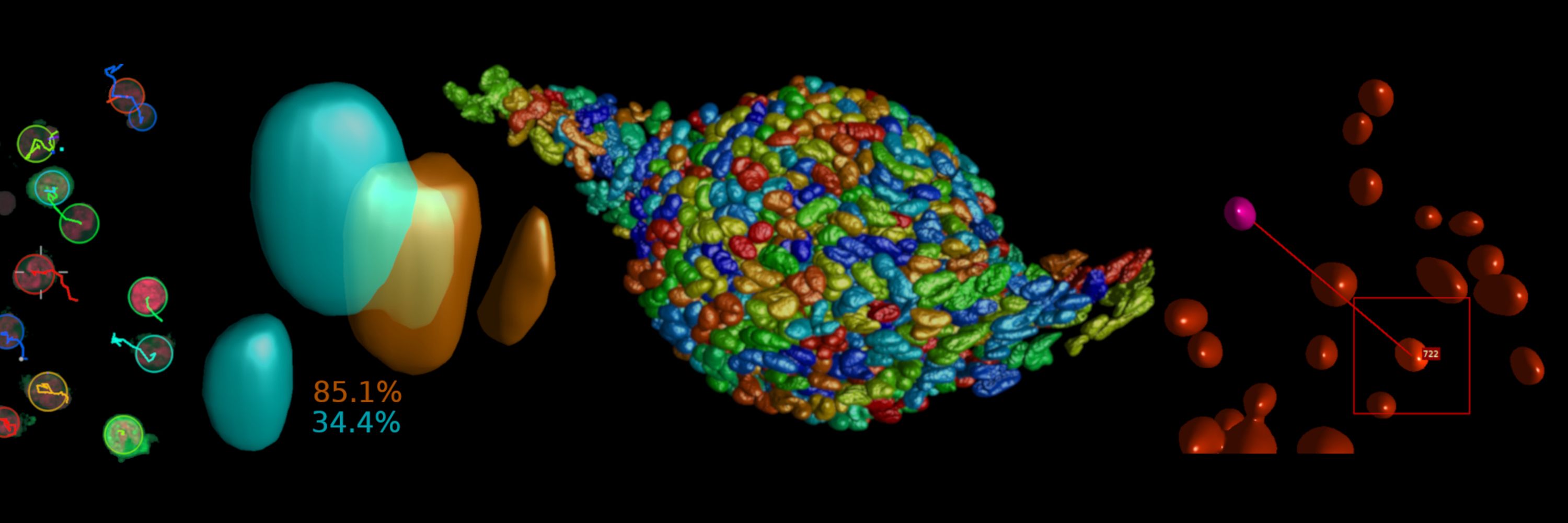
Microscopists, catch up anytime on our webinar on "Mastering Image Quality and Colocalization Analysis.
Visit: svi.nl/Huygens-Work...
Proper Acquisition -> QC -> True restoration -> Reliable Colocalization Analysis.
#imaging #microscopy #cellbiology #fluorescence #imageanalysis #colocalization
Visit: svi.nl/Huygens-Work...
Proper Acquisition -> QC -> True restoration -> Reliable Colocalization Analysis.
#imaging #microscopy #cellbiology #fluorescence #imageanalysis #colocalization
November 13, 2025 at 10:41 AM
Microscopists, catch up anytime on our webinar on "Mastering Image Quality and Colocalization Analysis.
Visit: svi.nl/Huygens-Work...
Proper Acquisition -> QC -> True restoration -> Reliable Colocalization Analysis.
#imaging #microscopy #cellbiology #fluorescence #imageanalysis #colocalization
Visit: svi.nl/Huygens-Work...
Proper Acquisition -> QC -> True restoration -> Reliable Colocalization Analysis.
#imaging #microscopy #cellbiology #fluorescence #imageanalysis #colocalization
Dear Imaging Experts: participate in this year's Huygens #Microscopy Image Contest with your most beautiful images and movies and win a laptop or SSD with a microscope image! See this website on how to participate: svi.nl/ImageContest
#ScienceArt #Microscopy #deconvolution #imageanalysis
#ScienceArt #Microscopy #deconvolution #imageanalysis

November 10, 2025 at 9:00 AM
Dear Imaging Experts: participate in this year's Huygens #Microscopy Image Contest with your most beautiful images and movies and win a laptop or SSD with a microscope image! See this website on how to participate: svi.nl/ImageContest
#ScienceArt #Microscopy #deconvolution #imageanalysis
#ScienceArt #Microscopy #deconvolution #imageanalysis
Master colocalization! Join our webinar Nov. 11 for multiple time zones: svi.nl/WebinarSched...
November 5, 2025 at 8:02 AM
Master colocalization! Join our webinar Nov. 11 for multiple time zones: svi.nl/WebinarSched...
“More than just Pearson” is one of the topics we address. Still a few hours left to subscribe for our FREE webinar on mastering Colocalization Analysis: svi.nl/Webinarinvit...
#imaging #microscopy #cellbiology #fluorescence #imageanalysis #colocalization
#imaging #microscopy #cellbiology #fluorescence #imageanalysis #colocalization

October 28, 2025 at 9:49 AM
“More than just Pearson” is one of the topics we address. Still a few hours left to subscribe for our FREE webinar on mastering Colocalization Analysis: svi.nl/Webinarinvit...
#imaging #microscopy #cellbiology #fluorescence #imageanalysis #colocalization
#imaging #microscopy #cellbiology #fluorescence #imageanalysis #colocalization
Join our open webinar on Mastering image quality and Colocalization Analysis: 👉 svi.nl/WebinarSched...
Colocalization analysis in microscopy might look simple at first — but once you dive in, the decisions you need to make can be challenging.
Join and learn how to attain the best possible results.
Colocalization analysis in microscopy might look simple at first — but once you dive in, the decisions you need to make can be challenging.
Join and learn how to attain the best possible results.
WebinarSchedule | Scientific Volume Imaging
Scientific Volume Imaging to provides reliable, high quality, easy to use image processing tools for scientists working in light microscopy. Together with a dedicated team in close contact with the in...
svi.nl
October 24, 2025 at 7:47 AM
Join our open webinar on Mastering image quality and Colocalization Analysis: 👉 svi.nl/WebinarSched...
Colocalization analysis in microscopy might look simple at first — but once you dive in, the decisions you need to make can be challenging.
Join and learn how to attain the best possible results.
Colocalization analysis in microscopy might look simple at first — but once you dive in, the decisions you need to make can be challenging.
Join and learn how to attain the best possible results.
Final week to subscribe for our FREE Webinar Series on "Mastering Colocalization Analysis"; From raw image to scientific results in minutes. Reserve your seat now!
svi.nl/webinarinvit...
#imaging #microscopy #cellbiology #fluorescence #imageanalysis #colocalization
svi.nl/webinarinvit...
#imaging #microscopy #cellbiology #fluorescence #imageanalysis #colocalization
October 22, 2025 at 11:32 AM
Final week to subscribe for our FREE Webinar Series on "Mastering Colocalization Analysis"; From raw image to scientific results in minutes. Reserve your seat now!
svi.nl/webinarinvit...
#imaging #microscopy #cellbiology #fluorescence #imageanalysis #colocalization
svi.nl/webinarinvit...
#imaging #microscopy #cellbiology #fluorescence #imageanalysis #colocalization
Happy #MicroscopyMonday! Learn how to view images that far exceed the RAM of your computer during our webinar tomorrow Oct. 21st at 9:30 or 1800 CEST: svi.nl/webinarinvit... and discover how the support for multi-resolution pyramid files enables you to stitch and visualize extremely large datasets!
WebinarInvitation | Scientific Volume Imaging
Scientific Volume Imaging to provides reliable, high quality, easy to use image processing tools for scientists working in light microscopy. Together with a dedicated team in close contact with the in...
svi.nl
October 20, 2025 at 9:19 AM
Happy #MicroscopyMonday! Learn how to view images that far exceed the RAM of your computer during our webinar tomorrow Oct. 21st at 9:30 or 1800 CEST: svi.nl/webinarinvit... and discover how the support for multi-resolution pyramid files enables you to stitch and visualize extremely large datasets!
Visiting @BioimagingNA #GBIxBINA 2025 in Montréal?
Let’s talk Image Quality Control for #colocalization and object analysis!
Look out for this T-shirt and stop by our poster — we’d love to chat.
In the meantime, test your own images (for free!) here 👉 userportal.svi.nl/Quality-Cont...
Let’s talk Image Quality Control for #colocalization and object analysis!
Look out for this T-shirt and stop by our poster — we’d love to chat.
In the meantime, test your own images (for free!) here 👉 userportal.svi.nl/Quality-Cont...

September 29, 2025 at 2:04 PM
Visiting @BioimagingNA #GBIxBINA 2025 in Montréal?
Let’s talk Image Quality Control for #colocalization and object analysis!
Look out for this T-shirt and stop by our poster — we’d love to chat.
In the meantime, test your own images (for free!) here 👉 userportal.svi.nl/Quality-Cont...
Let’s talk Image Quality Control for #colocalization and object analysis!
Look out for this T-shirt and stop by our poster — we’d love to chat.
In the meantime, test your own images (for free!) here 👉 userportal.svi.nl/Quality-Cont...
A Wormhole in our Universe?
Animation of a curled up C. elegans nematode imaged with a spinning disk confocal, deconvolved and rendered using the Huygens Software. C/o Igor Bonacossa Pereira, Hilliard Lab, Queensland Brain Institute.
#Neuroscience #microscopy #deconvolution #imageanalysis
Animation of a curled up C. elegans nematode imaged with a spinning disk confocal, deconvolved and rendered using the Huygens Software. C/o Igor Bonacossa Pereira, Hilliard Lab, Queensland Brain Institute.
#Neuroscience #microscopy #deconvolution #imageanalysis
September 19, 2025 at 10:48 AM
A Wormhole in our Universe?
Animation of a curled up C. elegans nematode imaged with a spinning disk confocal, deconvolved and rendered using the Huygens Software. C/o Igor Bonacossa Pereira, Hilliard Lab, Queensland Brain Institute.
#Neuroscience #microscopy #deconvolution #imageanalysis
Animation of a curled up C. elegans nematode imaged with a spinning disk confocal, deconvolved and rendered using the Huygens Software. C/o Igor Bonacossa Pereira, Hilliard Lab, Queensland Brain Institute.
#Neuroscience #microscopy #deconvolution #imageanalysis
#Mousebrain with neurons from the eyes. This cleared 870 GB tiled z-stack from a @zeiss_micro #LightSheet Z.1 was deconvolved, stitched, and vignetting corrected with Huygens.
Courtesy: Dominic Fillion, @ircm.bsky.social Keen to learn more? Join our webinar Sept 16th: svi.nl/webinarinvitation
Courtesy: Dominic Fillion, @ircm.bsky.social Keen to learn more? Join our webinar Sept 16th: svi.nl/webinarinvitation
September 11, 2025 at 7:47 AM
#Mousebrain with neurons from the eyes. This cleared 870 GB tiled z-stack from a @zeiss_micro #LightSheet Z.1 was deconvolved, stitched, and vignetting corrected with Huygens.
Courtesy: Dominic Fillion, @ircm.bsky.social Keen to learn more? Join our webinar Sept 16th: svi.nl/webinarinvitation
Courtesy: Dominic Fillion, @ircm.bsky.social Keen to learn more? Join our webinar Sept 16th: svi.nl/webinarinvitation
🔬 Exciting news!
SVI is now a proud partner of the @gdrimabio.bsky.social and will be present at #Mifobio2025!
We’re looking forward to this inspiring event — a great opportunity to connect and exchange ideas on Deconvolution, Image restoration, Analysis and Imaging quality control.
SVI is now a proud partner of the @gdrimabio.bsky.social and will be present at #Mifobio2025!
We’re looking forward to this inspiring event — a great opportunity to connect and exchange ideas on Deconvolution, Image restoration, Analysis and Imaging quality control.

September 9, 2025 at 7:52 AM
🔬 Exciting news!
SVI is now a proud partner of the @gdrimabio.bsky.social and will be present at #Mifobio2025!
We’re looking forward to this inspiring event — a great opportunity to connect and exchange ideas on Deconvolution, Image restoration, Analysis and Imaging quality control.
SVI is now a proud partner of the @gdrimabio.bsky.social and will be present at #Mifobio2025!
We’re looking forward to this inspiring event — a great opportunity to connect and exchange ideas on Deconvolution, Image restoration, Analysis and Imaging quality control.
Cellular spheroid of HeLa Kyoto cells. Left: 2 rotated @LaVision_BioTec UM views, deconvolved+fused+rendered with Huygens. Right: 1 view.
Join our Light Sheet webinar: svi.nl/Webinarinvitation Sept 16th.
Courtesy: Stylianos Didaskalou & Lito Karkaletsou of Prof. M. Koffa's lab -DUTH-MBG
Join our Light Sheet webinar: svi.nl/Webinarinvitation Sept 16th.
Courtesy: Stylianos Didaskalou & Lito Karkaletsou of Prof. M. Koffa's lab -DUTH-MBG

September 2, 2025 at 8:03 AM
Cellular spheroid of HeLa Kyoto cells. Left: 2 rotated @LaVision_BioTec UM views, deconvolved+fused+rendered with Huygens. Right: 1 view.
Join our Light Sheet webinar: svi.nl/Webinarinvitation Sept 16th.
Courtesy: Stylianos Didaskalou & Lito Karkaletsou of Prof. M. Koffa's lab -DUTH-MBG
Join our Light Sheet webinar: svi.nl/Webinarinvitation Sept 16th.
Courtesy: Stylianos Didaskalou & Lito Karkaletsou of Prof. M. Koffa's lab -DUTH-MBG
Reposted by SVI Huygens
Before vs After: Can you spot the difference in colocalization? Join our free webinar July 10th svi.nl/webinarinvitation on colocalization. Can't wait? doi.org/10.1093/mict...
#Microscopy #ImageRestoration #BioImageAnalysis #QuantitativeImaging #superresolution #AccurateColocalizationAnalysis
#Microscopy #ImageRestoration #BioImageAnalysis #QuantitativeImaging #superresolution #AccurateColocalizationAnalysis

July 3, 2025 at 6:44 AM
Before vs After: Can you spot the difference in colocalization? Join our free webinar July 10th svi.nl/webinarinvitation on colocalization. Can't wait? doi.org/10.1093/mict...
#Microscopy #ImageRestoration #BioImageAnalysis #QuantitativeImaging #superresolution #AccurateColocalizationAnalysis
#Microscopy #ImageRestoration #BioImageAnalysis #QuantitativeImaging #superresolution #AccurateColocalizationAnalysis
July 3, 2025 at 6:44 AM
Before vs After: Can you spot the difference in colocalization? Join our free webinar July 10th svi.nl/webinarinvitation on colocalization. Can't wait? doi.org/10.1093/mict...
#Microscopy #ImageRestoration #BioImageAnalysis #QuantitativeImaging #superresolution #AccurateColocalizationAnalysis
#Microscopy #ImageRestoration #BioImageAnalysis #QuantitativeImaging #superresolution #AccurateColocalizationAnalysis
🌟 Happy #FluorescenceFriday! 🌟
Before vs. After: Chromatic Aberration Correction – Can you spot the difference in colocalization?
Want to learn more? Sign up for our Webinar July 10th: svi.nl/webinarinvitation, or read this paper: academic.oup.com/mt/article/3...
#AccurateColocalizationAnalysis
Before vs. After: Chromatic Aberration Correction – Can you spot the difference in colocalization?
Want to learn more? Sign up for our Webinar July 10th: svi.nl/webinarinvitation, or read this paper: academic.oup.com/mt/article/3...
#AccurateColocalizationAnalysis

June 27, 2025 at 8:10 AM
🌟 Happy #FluorescenceFriday! 🌟
Before vs. After: Chromatic Aberration Correction – Can you spot the difference in colocalization?
Want to learn more? Sign up for our Webinar July 10th: svi.nl/webinarinvitation, or read this paper: academic.oup.com/mt/article/3...
#AccurateColocalizationAnalysis
Before vs. After: Chromatic Aberration Correction – Can you spot the difference in colocalization?
Want to learn more? Sign up for our Webinar July 10th: svi.nl/webinarinvitation, or read this paper: academic.oup.com/mt/article/3...
#AccurateColocalizationAnalysis