


Manon Janet-Maitre
@manonjanet.bsky.social
Postdoc in the Feldman lab, WashU in St Louis, USA - PhD in the Attree lab, Grenoble Alpes University, France
Crazy bacteria lover 🧫🦠🥼
Crazy bacteria lover 🧫🦠🥼
Finally out! Right on time for the holidays 🎄🎁
Big thanks to Nick Scott, @giseladivenanzio.bsky.social, Clay and Mario @feldmanlab.bsky.social who made it happen!
www.pnas.org/doi/10.1073/...
Big thanks to Nick Scott, @giseladivenanzio.bsky.social, Clay and Mario @feldmanlab.bsky.social who made it happen!
www.pnas.org/doi/10.1073/...
December 9, 2025 at 7:40 PM
Finally out! Right on time for the holidays 🎄🎁
Big thanks to Nick Scott, @giseladivenanzio.bsky.social, Clay and Mario @feldmanlab.bsky.social who made it happen!
www.pnas.org/doi/10.1073/...
Big thanks to Nick Scott, @giseladivenanzio.bsky.social, Clay and Mario @feldmanlab.bsky.social who made it happen!
www.pnas.org/doi/10.1073/...
Happy to share that our paper on the complement resistome of Pseudomonas aeruginosa is finally out in I&I! This work was done during my PhD in the lab of @inaattree.bsky.social and made possible by my amazing co-authors and colleagues 🥳
You can check it out here: journals.asm.org/doi/10.1128/...
You can check it out here: journals.asm.org/doi/10.1128/...
Strain-specific variation in the complement resistome of Pseudomonas aeruginosa | Infection and Immunity
Bacterial bloodstream infections (BSIs) represent a huge burden for modern healthcare. BSIs caused by Pseudomonas aeruginosa exhibit the highest mortality rate (1, 2). This leading nosocomial pathogen causes various primary infections, such as burn wounds, pulmonary and urinary tract infections (3). Using its arsenal of virulence factors, it is able to breach both epithelial and endothelial barriers ([4–7], reviewed in [8]). Once in the bloodstream, P. aeruginosa faces the host immune response. In a previous study, we showed the complement system to be the main innate immune component responsible for the elimination of P. aeruginosa in the blood (9). The complement system is an enzymatic cascade of more than 30 proteins, whose activation results in the formation of the membrane attack complex (MAC) in the outer membrane of the target pathogen. Complement-mediated killing happens through three main steps including complement activation, assembly of the C5b-9 MAC on the outer membrane, and penetration of the MAC into the bilayer, eventually leading to bacterial lysis (10, 11). P. aeruginosa evolved strategies to evade those three steps. The secretion of proteases including AprA and LasB, cleaving C1, C2, and C3 or the secretion of ecotin, a protease inhibitor, leads to the blockade of complement activation (12–14). P. aeruginosa can also recruit host complement inhibitors to its surface, notably through the exposure of the elongation factor Tuf or the dihydrolipoamide dehydrogenase Lpd ([15, 16], reviewed in [8]). These immune evasion proteins can recruit the Factor H family of complement regulators, resulting in C3b degradation and preventing downstream proteolysis cascade. Finally, P. aeruginosa can modify its surface by producing exopolysaccharides (EPSs) such as alginate. When acylated, alginates decrease bacterial opsonization by the C3b molecule (17, 18).
journals.asm.org
August 13, 2025 at 1:51 PM
Happy to share that our paper on the complement resistome of Pseudomonas aeruginosa is finally out in I&I! This work was done during my PhD in the lab of @inaattree.bsky.social and made possible by my amazing co-authors and colleagues 🥳
You can check it out here: journals.asm.org/doi/10.1128/...
You can check it out here: journals.asm.org/doi/10.1128/...
This is my first bluesky post!
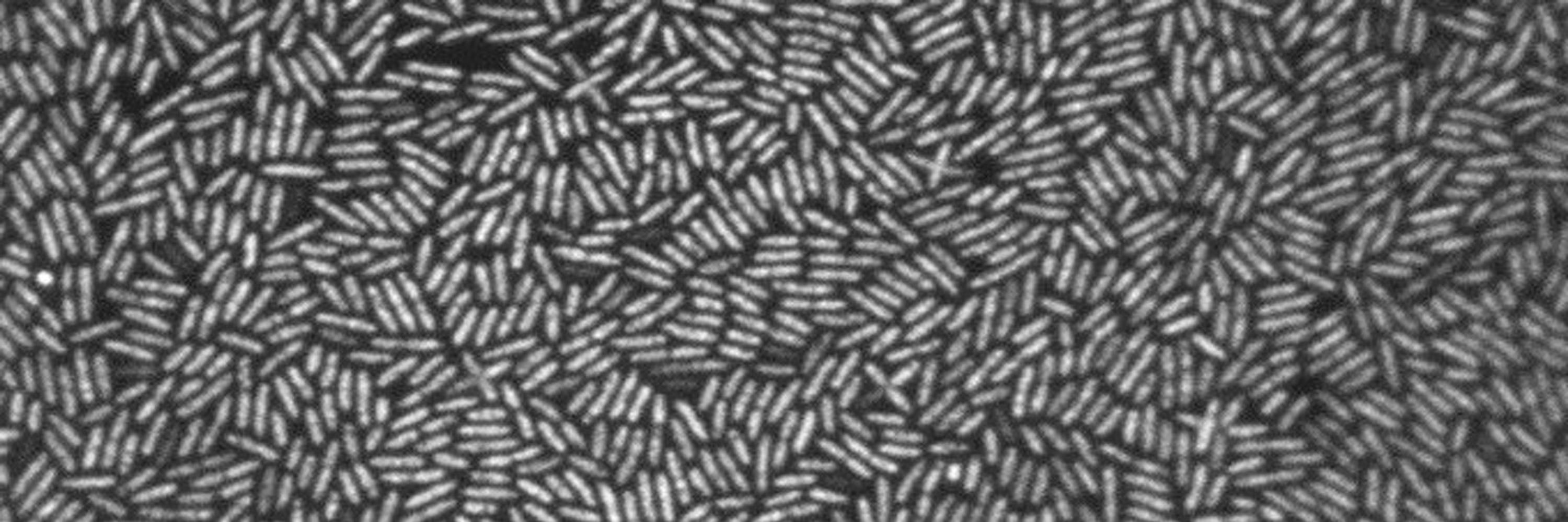
Excited to share this project I've been working on in the Feldman lab!🥳
We show that intracellular A. baumannii acts as a reservoir in mouse lung infection and characterize the interaction of A. baumannii with the macrophage using dual proteomics.
Excited to share this project I've been working on in the Feldman lab!🥳
We show that intracellular A. baumannii acts as a reservoir in mouse lung infection and characterize the interaction of A. baumannii with the macrophage using dual proteomics.

Yes! @manonjanet.bsky.social preprint is out! We show using BALF transfer that intracelular Acinetobacter baumannii can seed an infection in a susceptible mouse, suggesting that intracellular bacteria may be reservoir for recurrent infections
nam10.safelinks.protection.outlook.com?url=https%3A...
nam10.safelinks.protection.outlook.com?url=https%3A...
nam10.safelinks.protection.outlook.com
April 30, 2025 at 6:43 PM
This is my first bluesky post!
Excited to share this project I've been working on in the Feldman lab!🥳
We show that intracellular A. baumannii acts as a reservoir in mouse lung infection and characterize the interaction of A. baumannii with the macrophage using dual proteomics.
Excited to share this project I've been working on in the Feldman lab!🥳
We show that intracellular A. baumannii acts as a reservoir in mouse lung infection and characterize the interaction of A. baumannii with the macrophage using dual proteomics.