


Vaibhav Mahajan
@vaibhavmahajan.bsky.social
Ph.D. student with Anna Taubenberger, BIOTEC, TU Dresden | M.Res. with Buzz Baum, MRC LMCB, UCL | Interested in mechanics, 3D systems, evolution and all of cell biology
Han et al. had proposed that these volume changes during invasion are due to a compressive stress gradient (from centre to periphery). This drives intracellular flow of water between cells, through gap junctions. We do not see any gradient of volume in our spheroids (like most people in the field).

October 5, 2025 at 3:11 PM
Han et al. had proposed that these volume changes during invasion are due to a compressive stress gradient (from centre to periphery). This drives intracellular flow of water between cells, through gap junctions. We do not see any gradient of volume in our spheroids (like most people in the field).
Here we had a single cell becoming a multicellular entity. In invasion, it is the opposite - a multicellular entity gives rise to 'single' cells. We (along with Han et al. 2020 Nat Phy) see the same trend in this case. Invading cells are bigger compared to cells at the centre of the spheroid.


October 5, 2025 at 3:11 PM
Here we had a single cell becoming a multicellular entity. In invasion, it is the opposite - a multicellular entity gives rise to 'single' cells. We (along with Han et al. 2020 Nat Phy) see the same trend in this case. Invading cells are bigger compared to cells at the centre of the spheroid.
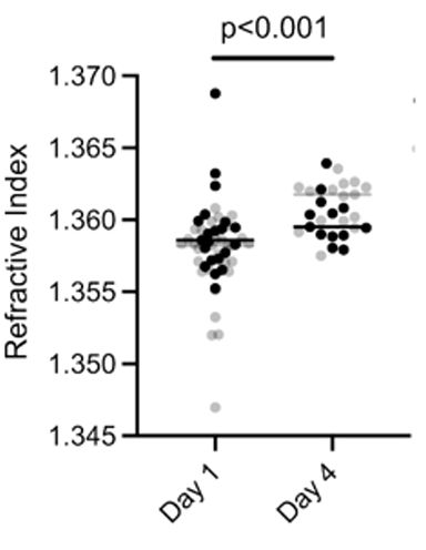
This was also suggested by looking at the refractive index of cells. RI scales with the mass density of cells. We observed higher RI for multicellular structures compared to single cells. So when cells were released from this context, they osmotically balanced themselves and increased in volume.

October 5, 2025 at 3:11 PM
This was also suggested by looking at the refractive index of cells. RI scales with the mass density of cells. We observed higher RI for multicellular structures compared to single cells. So when cells were released from this context, they osmotically balanced themselves and increased in volume.
We then released single cells at different timepoints (by breaking down the gel and the clusters/spheroids) and looked at their nuclear volumes. We observed that when cells were released from a multicellular context, they (probably) took in water and increased their nuclear volume.


October 5, 2025 at 3:11 PM
We then released single cells at different timepoints (by breaking down the gel and the clusters/spheroids) and looked at their nuclear volumes. We observed that when cells were released from a multicellular context, they (probably) took in water and increased their nuclear volume.
By blocking cells in S/G2 phase with a CDK1 inhibitor, we were able to rescue the nuclear volumes of small clusters. But this increased nuclear volume still did not reach the single cell level. This hinted that another mechanism was at play.


October 5, 2025 at 3:11 PM
By blocking cells in S/G2 phase with a CDK1 inhibitor, we were able to rescue the nuclear volumes of small clusters. But this increased nuclear volume still did not reach the single cell level. This hinted that another mechanism was at play.
As single cells formed these multicellular structures, there was an accumulation of smaller G1 cells over time. Light sheet microscopy allowed us to show that this was due to prolongation of the G1 phase of the cell cycle, while S/G2 phase length did not change much over time.


October 5, 2025 at 3:11 PM
As single cells formed these multicellular structures, there was an accumulation of smaller G1 cells over time. Light sheet microscopy allowed us to show that this was due to prolongation of the G1 phase of the cell cycle, while S/G2 phase length did not change much over time.
Then why are the nuclear volumes decreasing? We did some coarse timelapse microscopy to tackle that. We saw that cells were growing over the cell cycle and could become as big as their mother cell. So, there was no inhibition of growth in this system. But this hinted at us to look at the cell cycle.


October 5, 2025 at 3:11 PM
Then why are the nuclear volumes decreasing? We did some coarse timelapse microscopy to tackle that. We saw that cells were growing over the cell cycle and could become as big as their mother cell. So, there was no inhibition of growth in this system. But this hinted at us to look at the cell cycle.
You might guess that nuclear volumes might decrease due to compressive stress on the encapsulated spheroids. We tested that using the gel system. By changing the degradability and stiffness of the gel, we can alter the compressive stress on the spheroids. But that did not affect the nuclear volumes.


October 5, 2025 at 3:11 PM
You might guess that nuclear volumes might decrease due to compressive stress on the encapsulated spheroids. We tested that using the gel system. By changing the degradability and stiffness of the gel, we can alter the compressive stress on the spheroids. But that did not affect the nuclear volumes.
Our model system - single cells in 3D hydrogels that proliferated to form clonal spheroids. We were surprised to see drastic reductions in nuclear volumes upon nuclear segmentation in 3D. Below the data is for MCF-7 but we see this for multiple cell lines - PANC1, MCF10A, HeLa.


October 5, 2025 at 3:11 PM
Our model system - single cells in 3D hydrogels that proliferated to form clonal spheroids. We were surprised to see drastic reductions in nuclear volumes upon nuclear segmentation in 3D. Below the data is for MCF-7 but we see this for multiple cell lines - PANC1, MCF10A, HeLa.
I wrote this some time back - ‘PhD is for training’
December 1, 2024 at 4:59 PM
I wrote this some time back - ‘PhD is for training’