


Danielle Grotjahn
@nanigrotjahn.bsky.social
Associate prof @ Scripps Research
grotjahnlab.org
#teamtomo #mitochondriac
grotjahnlab.org
#teamtomo #mitochondriac
Pinned

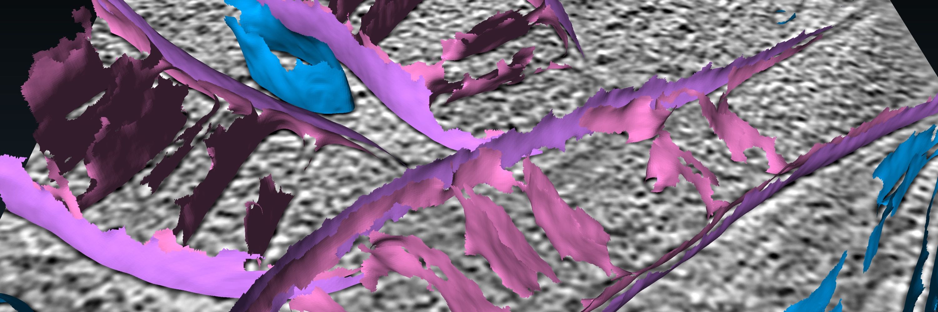
Surface morphometrics reveals local membrane thickness variation in organellar subcompartments https://www.biorxiv.org/content/10.1101/2025.04.30.651574v1
🚨New preprint!🚨 #teamtomo
We expanded Surface Morphometrics to quantify membrane thickness from cryo-ET—revealing local variation across organelles.
Led by the lab’s first grad student, @mmedina300kv.bsky.social (defending Monday! 🍾) w/ @attychang.bsky.social @hamid13r.bsky.social & @tomo.science
We expanded Surface Morphometrics to quantify membrane thickness from cryo-ET—revealing local variation across organelles.
Led by the lab’s first grad student, @mmedina300kv.bsky.social (defending Monday! 🍾) w/ @attychang.bsky.social @hamid13r.bsky.social & @tomo.science
Reposted by Danielle Grotjahn
Exciting !

BRET-Based Mitochondrial Subcompartment Localization Biosensors https://www.biorxiv.org/content/10.1101/2025.10.27.684728v1
October 28, 2025 at 9:13 PM
Exciting !
Reposted by Danielle Grotjahn
Thank you to all of our plenary speakers for sharing your incredible research with us at #ASBMB2025! @nanigrotjahn.bsky.social @ajknights7.bsky.social Michelle Haber and Randal Halfmann. We have loved having you all in Brisbane and hearing about your labs awesome work! 🦠🧬🧪🧫🔬


October 1, 2025 at 2:09 AM
Thank you to all of our plenary speakers for sharing your incredible research with us at #ASBMB2025! @nanigrotjahn.bsky.social @ajknights7.bsky.social Michelle Haber and Randal Halfmann. We have loved having you all in Brisbane and hearing about your labs awesome work! 🦠🧬🧪🧫🔬
Reposted by Danielle Grotjahn
Happy to announce "Technical Innovations Using #SerialEM: A Community Meeting", a new format of hybrid meeting David Mastronarde and I are organizing in April 2026. For more info, a preliminary list of speakers, pre-registration etc. visit
www.nexperion.net/semcm2026
www.nexperion.net/semcm2026

September 23, 2025 at 10:42 AM
Happy to announce "Technical Innovations Using #SerialEM: A Community Meeting", a new format of hybrid meeting David Mastronarde and I are organizing in April 2026. For more info, a preliminary list of speakers, pre-registration etc. visit
www.nexperion.net/semcm2026
www.nexperion.net/semcm2026
Reposted by Danielle Grotjahn
What an inspiring few days at the #WEHeraeusSeminar! Great science, lively discussions, and lots of fun with the amazing community of #cryoEM, #StructuralBiology & #Microscopy. Huge thanks to everyone!

Big thanks to @arnemoeller.bsky.social , @djanuliene.bsky.social, all speakers, and everyone who made the #WEHeraeusSeminar - Cryo-EM on the Move such an amazing event! It was a blast!
#cryoEM #StructuralBiology #Microscopy
#cryoEM #StructuralBiology #Microscopy

September 17, 2025 at 3:32 PM
What an inspiring few days at the #WEHeraeusSeminar! Great science, lively discussions, and lots of fun with the amazing community of #cryoEM, #StructuralBiology & #Microscopy. Huge thanks to everyone!
Reposted by Danielle Grotjahn
We all love colours, especially, when they can be generated automatically! Fantastic talk by Danielle Grotjahn @nanigrotjahn.bsky.social linking method development, structural biology and cell biology! #WEHeraeusSeminar #cryoEM #StructuralBiology #Microscopy @arnemoeller.bsky.social

September 16, 2025 at 3:28 PM
We all love colours, especially, when they can be generated automatically! Fantastic talk by Danielle Grotjahn @nanigrotjahn.bsky.social linking method development, structural biology and cell biology! #WEHeraeusSeminar #cryoEM #StructuralBiology #Microscopy @arnemoeller.bsky.social
Excited to travel to Germany 🇩🇪 for the WE-Heraeus-Seminar: Cryo‐EM on the Move: New Applications and Challenges! ❄️🔬🧊
I look forward to hearing what everyone is cooking up to make cryoEM better 📈 faster 🏃♀️ stronger 💪 😎
www.we-heraeus-stiftung.de/veranstaltun...
I look forward to hearing what everyone is cooking up to make cryoEM better 📈 faster 🏃♀️ stronger 💪 😎
www.we-heraeus-stiftung.de/veranstaltun...
Wilhelm und Else Heraeus-Stiftung: Main
www.we-heraeus-stiftung.de
September 12, 2025 at 6:29 PM
Excited to travel to Germany 🇩🇪 for the WE-Heraeus-Seminar: Cryo‐EM on the Move: New Applications and Challenges! ❄️🔬🧊
I look forward to hearing what everyone is cooking up to make cryoEM better 📈 faster 🏃♀️ stronger 💪 😎
www.we-heraeus-stiftung.de/veranstaltun...
I look forward to hearing what everyone is cooking up to make cryoEM better 📈 faster 🏃♀️ stronger 💪 😎
www.we-heraeus-stiftung.de/veranstaltun...
🚨 #teamtomo!🚨
Did you know there's a dedicated cryoEM/ET Mini-Symposium at the upcoming ASCB/EMBO meeting in Philly (Dec 6–10)??
Did you know there's a dedicated cryoEM/ET Mini-Symposium at the upcoming ASCB/EMBO meeting in Philly (Dec 6–10)??
August 20, 2025 at 12:18 AM
🚨 #teamtomo!🚨
Did you know there's a dedicated cryoEM/ET Mini-Symposium at the upcoming ASCB/EMBO meeting in Philly (Dec 6–10)??
Did you know there's a dedicated cryoEM/ET Mini-Symposium at the upcoming ASCB/EMBO meeting in Philly (Dec 6–10)??
Ending the week on a high note in SLC with a visit to @uofubiochem.bsky.social after an inspiring M&M 2025 meeting!

On Thurs July 31st, Dr. Danielle Grotjahn @nanigrotjahn.bsky.social (Scripps) will give a talk entitled "Lessons from mitochondria on thriving under stress." 12pm in HSEB Alumni Hall!

July 29, 2025 at 7:01 PM
Ending the week on a high note in SLC with a visit to @uofubiochem.bsky.social after an inspiring M&M 2025 meeting!
I have lots of opinions—but for your sake, I’ve limited them to segmentation in cryo-ET for this article 😉
www.sciencedirect.com/science/arti...
#teamtomo
www.sciencedirect.com/science/arti...
#teamtomo

Segmenting cryo-electron tomography data: Extracting models from cellular landscapes
Cryo-electron tomography provides an unprecedented view of cellular architecture, yet extracting meaningful biological insights remains challenging. S…
www.sciencedirect.com
July 11, 2025 at 12:46 AM
I have lots of opinions—but for your sake, I’ve limited them to segmentation in cryo-ET for this article 😉
www.sciencedirect.com/science/arti...
#teamtomo
www.sciencedirect.com/science/arti...
#teamtomo
Reposted by Danielle Grotjahn
Happy to say that my Current Opinion in Structural Biology article came out today. I hope it helps to guide to you in using cryo-ET to make new biological discoveries!
www.sciencedirect.com/science/arti...
www.sciencedirect.com/science/arti...
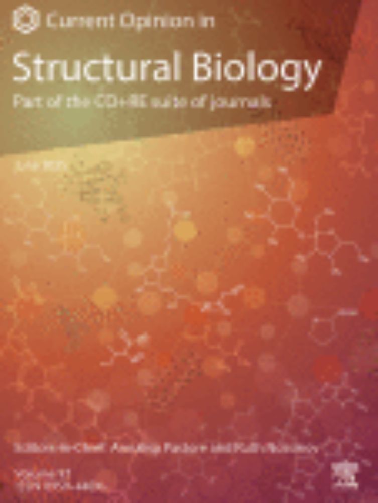
A practical look at cryo-electron tomography image processing: Key considerations for new biological discoveries
Cryo-electron tomography (cryo-ET) enables 3D visualization of complex biological environments without the need for purification, thereby preserving t…
www.sciencedirect.com
July 8, 2025 at 9:44 PM
Happy to say that my Current Opinion in Structural Biology article came out today. I hope it helps to guide to you in using cryo-ET to make new biological discoveries!
www.sciencedirect.com/science/arti...
www.sciencedirect.com/science/arti...
Reposted by Danielle Grotjahn

New work from the lab (w/ @preronabora.bsky.social @mashiatzaman.bsky.social @mitomorph.bsky.social @nanigrotjahn.bsky.social) identifying a compound that activates OMA1-DELE1-HRI signaling to induce mitochondrial remodeling in MFN2-deficient cells. Check it out 👇
www.biorxiv.org/content/10.1...
www.biorxiv.org/content/10.1...
Drug Repurposing Screen Identifies an HRI Activating Compound that Promotes Adaptive Mitochondrial Remodeling in MFN2-deficient Cells
Pathogenic variants in the mitochondrial outer membrane GTPase MFN2 cause the peripheral neuropathy Charcot-Marie-Tooth Type 2A (CMT2A). These mutations disrupt MFN2-dependent regulation of diverse as...
www.biorxiv.org
June 27, 2025 at 3:52 PM
New work from the lab (w/ @preronabora.bsky.social @mashiatzaman.bsky.social @mitomorph.bsky.social @nanigrotjahn.bsky.social) identifying a compound that activates OMA1-DELE1-HRI signaling to induce mitochondrial remodeling in MFN2-deficient cells. Check it out 👇
www.biorxiv.org/content/10.1...
www.biorxiv.org/content/10.1...
Reposted by Danielle Grotjahn
I spoke with Jade Hindmon, host of @kpbssandiego.bsky.social Midday Edition, about the consequences of the current administration’s cuts to science funding. Please share to help spread the word and raise awareness.
www.kpbs.org/podcasts/kpb...
www.kpbs.org/podcasts/kpb...
Nobel laureate on how Trump's cuts are hurting science
Neuroscientist, biologist and 2021 Nobel Prize winner Ardem Patapoutian joined Midday Edition to talk about how the Trump administration's effort to cut back on science spending is impacting researche...
www.kpbs.org
June 19, 2025 at 12:27 AM
I spoke with Jade Hindmon, host of @kpbssandiego.bsky.social Midday Edition, about the consequences of the current administration’s cuts to science funding. Please share to help spread the word and raise awareness.
www.kpbs.org/podcasts/kpb...
www.kpbs.org/podcasts/kpb...
Reposted by Danielle Grotjahn
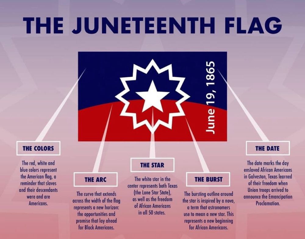
As we approach the week leading up to Juneteenth, it’s important to recognize the flag and symbols around the day, as well as taking the opportunity to renew the call for lineage-based reparations for not only chattel slavery, but the harms inflicted upon Black Americans post-emancipation.
June 15, 2025 at 4:11 PM
As we approach the week leading up to Juneteenth, it’s important to recognize the flag and symbols around the day, as well as taking the opportunity to renew the call for lineage-based reparations for not only chattel slavery, but the harms inflicted upon Black Americans post-emancipation.
It’s been a big week for the Grotjahn Lab—our very first graduate student, @mmedina300kv.bsky.social, successfully defended her PhD thesis! 🎉 So proud of everything she’s accomplished and grateful for the light she’s brought to our lab from day one ☀️⚡
May 10, 2025 at 1:11 AM
It’s been a big week for the Grotjahn Lab—our very first graduate student, @mmedina300kv.bsky.social, successfully defended her PhD thesis! 🎉 So proud of everything she’s accomplished and grateful for the light she’s brought to our lab from day one ☀️⚡
Tying self-worth to academic success, only to ask years later, "who am I, really?"
Grateful @frezzalab.bsky.social took the time to capture this tension so beautifully in this article.
Grateful @frezzalab.bsky.social took the time to capture this tension so beautifully in this article.
I'd like to share this short piece on a topic I hope you will find useful. It is the result of a personl journey and many discussions with friends and peers. Let me know your thoughts.
network.febs.org/posts/the-pr...
network.febs.org/posts/the-pr...
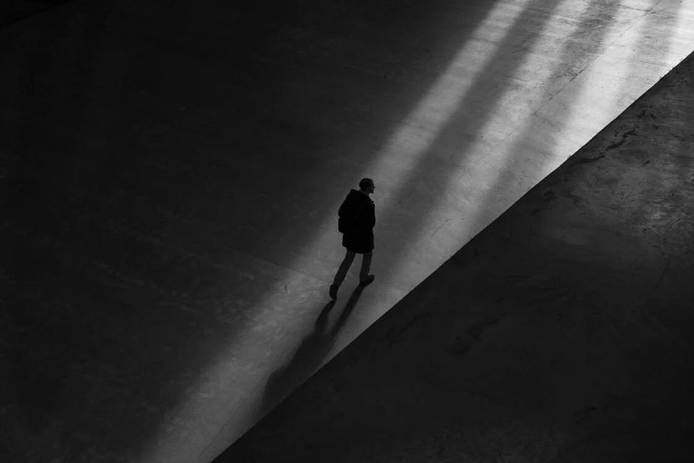
The Professional Self: The Enemy Inside
network.febs.org
May 9, 2025 at 7:46 PM
Tying self-worth to academic success, only to ask years later, "who am I, really?"
Grateful @frezzalab.bsky.social took the time to capture this tension so beautifully in this article.
Grateful @frezzalab.bsky.social took the time to capture this tension so beautifully in this article.
Reposted by Danielle Grotjahn
So excited for this work to be out!! Thanks to @attychang.bsky.social, @hamid13r.bsky.social, blue sky less Daniel Fuentes, @nanigrotjahn.bsky.social and @tomo.science!!!
🚨New preprint!🚨 #teamtomo
We expanded Surface Morphometrics to quantify membrane thickness from cryo-ET—revealing local variation across organelles.
Led by the lab’s first grad student, @mmedina300kv.bsky.social (defending Monday! 🍾) w/ @attychang.bsky.social @hamid13r.bsky.social & @tomo.science
We expanded Surface Morphometrics to quantify membrane thickness from cryo-ET—revealing local variation across organelles.
Led by the lab’s first grad student, @mmedina300kv.bsky.social (defending Monday! 🍾) w/ @attychang.bsky.social @hamid13r.bsky.social & @tomo.science
Surface morphometrics reveals local membrane thickness variation in organellar subcompartments https://www.biorxiv.org/content/10.1101/2025.04.30.651574v1
May 1, 2025 at 8:54 PM
So excited for this work to be out!! Thanks to @attychang.bsky.social, @hamid13r.bsky.social, blue sky less Daniel Fuentes, @nanigrotjahn.bsky.social and @tomo.science!!!
🚨New preprint!🚨 #teamtomo
We expanded Surface Morphometrics to quantify membrane thickness from cryo-ET—revealing local variation across organelles.
Led by the lab’s first grad student, @mmedina300kv.bsky.social (defending Monday! 🍾) w/ @attychang.bsky.social @hamid13r.bsky.social & @tomo.science
We expanded Surface Morphometrics to quantify membrane thickness from cryo-ET—revealing local variation across organelles.
Led by the lab’s first grad student, @mmedina300kv.bsky.social (defending Monday! 🍾) w/ @attychang.bsky.social @hamid13r.bsky.social & @tomo.science
Surface morphometrics reveals local membrane thickness variation in organellar subcompartments https://www.biorxiv.org/content/10.1101/2025.04.30.651574v1
May 1, 2025 at 7:42 PM
🚨New preprint!🚨 #teamtomo
We expanded Surface Morphometrics to quantify membrane thickness from cryo-ET—revealing local variation across organelles.
Led by the lab’s first grad student, @mmedina300kv.bsky.social (defending Monday! 🍾) w/ @attychang.bsky.social @hamid13r.bsky.social & @tomo.science
We expanded Surface Morphometrics to quantify membrane thickness from cryo-ET—revealing local variation across organelles.
Led by the lab’s first grad student, @mmedina300kv.bsky.social (defending Monday! 🍾) w/ @attychang.bsky.social @hamid13r.bsky.social & @tomo.science
Reposted by Danielle Grotjahn
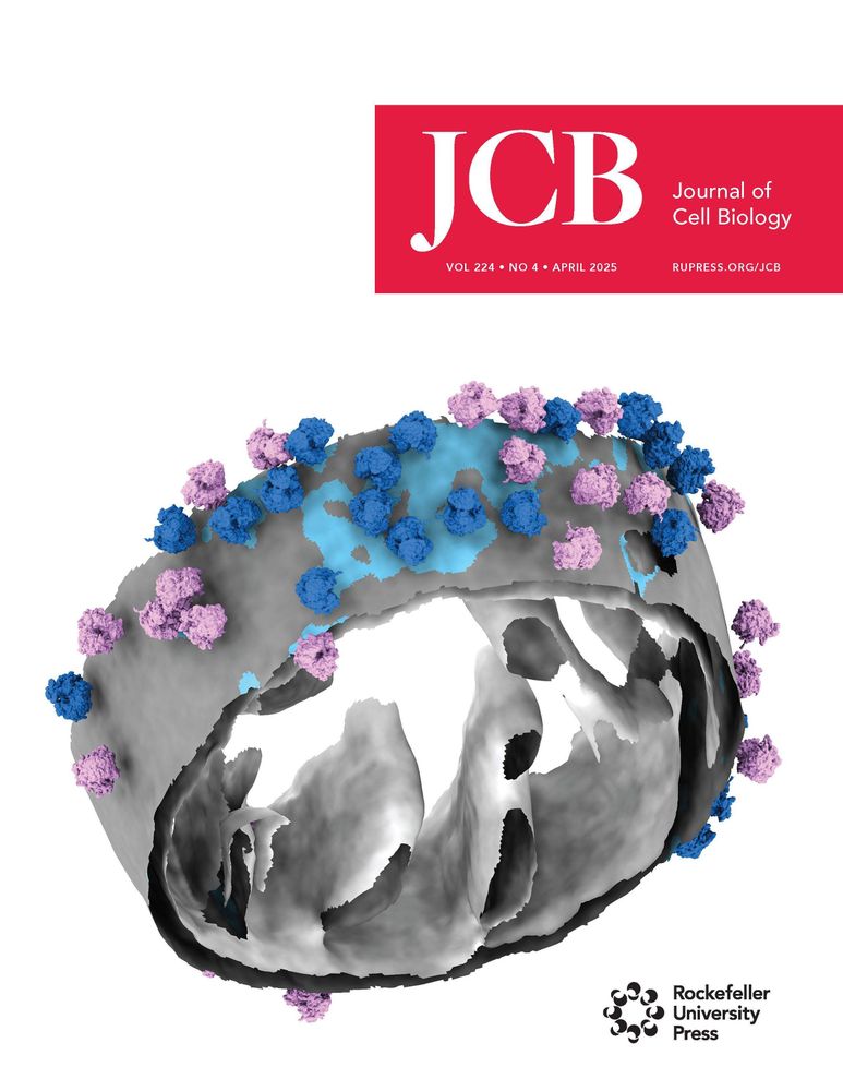
Feeling so honored and thrilled to see our work make it to the cover—what a milestone in my career! I can’t wait to receive the poster from JCB🤩
Our April issue is here! rupress.org/jcb/issue/22...
The cover shows a segmented model of cytoplasmic #ribosomes associated with mitochondrial membrane in a Saccharomyces cerevisiae cell. From Ya-Ting Chang, @nanigrotjahn.bsky.social and colleagues (doi.org/10.1083/jcb....)
The cover shows a segmented model of cytoplasmic #ribosomes associated with mitochondrial membrane in a Saccharomyces cerevisiae cell. From Ya-Ting Chang, @nanigrotjahn.bsky.social and colleagues (doi.org/10.1083/jcb....)

April 10, 2025 at 5:18 AM
Feeling so honored and thrilled to see our work make it to the cover—what a milestone in my career! I can’t wait to receive the poster from JCB🤩
Reposted by Danielle Grotjahn

@jcb.org's April issue is here! rupress.org/jcb/issue/22...
The cover shows a segmented model of cytoplasmic #ribosomes associated with mitochondrial membrane in a Saccharomyces cerevisiae cell. From Ya-Ting Chang, @nanigrotjahn.bsky.social and colleagues (doi.org/10.1083/jcb....)
The cover shows a segmented model of cytoplasmic #ribosomes associated with mitochondrial membrane in a Saccharomyces cerevisiae cell. From Ya-Ting Chang, @nanigrotjahn.bsky.social and colleagues (doi.org/10.1083/jcb....)
April 7, 2025 at 2:20 PM
@jcb.org's April issue is here! rupress.org/jcb/issue/22...
The cover shows a segmented model of cytoplasmic #ribosomes associated with mitochondrial membrane in a Saccharomyces cerevisiae cell. From Ya-Ting Chang, @nanigrotjahn.bsky.social and colleagues (doi.org/10.1083/jcb....)
The cover shows a segmented model of cytoplasmic #ribosomes associated with mitochondrial membrane in a Saccharomyces cerevisiae cell. From Ya-Ting Chang, @nanigrotjahn.bsky.social and colleagues (doi.org/10.1083/jcb....)
I know print journals aren't exactly on everyone's radar these days, but we’re still geeking out that our latest study—led by the brilliant @attychang.bsky.social—landed the cover of the April issue of JCB! 🤩
@zidlab.bsky.social @tomo.science @hamid13r.bsky.social
@zidlab.bsky.social @tomo.science @hamid13r.bsky.social
Our April issue is here! rupress.org/jcb/issue/22...
The cover shows a segmented model of cytoplasmic #ribosomes associated with mitochondrial membrane in a Saccharomyces cerevisiae cell. From Ya-Ting Chang, @nanigrotjahn.bsky.social and colleagues (doi.org/10.1083/jcb....)
The cover shows a segmented model of cytoplasmic #ribosomes associated with mitochondrial membrane in a Saccharomyces cerevisiae cell. From Ya-Ting Chang, @nanigrotjahn.bsky.social and colleagues (doi.org/10.1083/jcb....)
April 9, 2025 at 10:35 PM
I know print journals aren't exactly on everyone's radar these days, but we’re still geeking out that our latest study—led by the brilliant @attychang.bsky.social—landed the cover of the April issue of JCB! 🤩
@zidlab.bsky.social @tomo.science @hamid13r.bsky.social
@zidlab.bsky.social @tomo.science @hamid13r.bsky.social
Reposted by Danielle Grotjahn

Cytoplasmic #ribosomes on #mitochondria alter the local membrane environment for protein import, say Ya-Ting Chang, Danielle Grotjahn (@nanigrotjahn.bsky.social) @scripps.edu and colleagues: rupress.org/jcb/article/...
#ProteinHomeostasis #Organelles #StructuralBiology #CryoET
#ProteinHomeostasis #Organelles #StructuralBiology #CryoET
March 13, 2025 at 8:32 PM
Cytoplasmic #ribosomes on #mitochondria alter the local membrane environment for protein import, say Ya-Ting Chang, Danielle Grotjahn (@nanigrotjahn.bsky.social) @scripps.edu and colleagues: rupress.org/jcb/article/...
#ProteinHomeostasis #Organelles #StructuralBiology #CryoET
#ProteinHomeostasis #Organelles #StructuralBiology #CryoET
Reposted by Danielle Grotjahn

Do you LOVE organelles??
Well I am THRILLED to announce that Nellie has been published in @naturemethods.bsky.social!
Nellie a fully automated pipeline for organelle segmentation, tracking, and hierarchical feature extraction in 2D, 3D, timelapse, multichannel live-cell microscopy
🧵1/N
Well I am THRILLED to announce that Nellie has been published in @naturemethods.bsky.social!
Nellie a fully automated pipeline for organelle segmentation, tracking, and hierarchical feature extraction in 2D, 3D, timelapse, multichannel live-cell microscopy
🧵1/N
February 27, 2025 at 8:13 PM
Do you LOVE organelles??
Well I am THRILLED to announce that Nellie has been published in @naturemethods.bsky.social!
Nellie a fully automated pipeline for organelle segmentation, tracking, and hierarchical feature extraction in 2D, 3D, timelapse, multichannel live-cell microscopy
🧵1/N
Well I am THRILLED to announce that Nellie has been published in @naturemethods.bsky.social!
Nellie a fully automated pipeline for organelle segmentation, tracking, and hierarchical feature extraction in 2D, 3D, timelapse, multichannel live-cell microscopy
🧵1/N
Reposted by Danielle Grotjahn
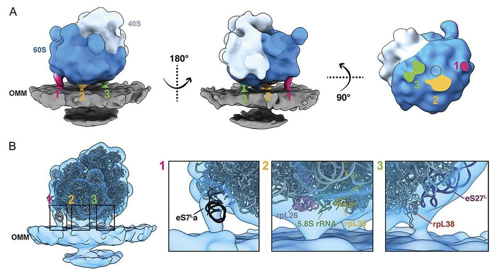
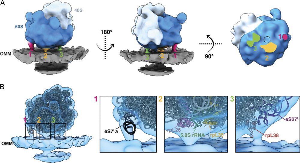
Ya-Ting Chang, @nanigrotjahn.bsky.social et al. @scripps.edu present a membrane-guided approach for identifying a subset of cytoplasmic #ribosomes oriented for protein import on the surface of #mitochondria in budding yeast using #cryoET: rupress.org/jcb/article/...
March 6, 2025 at 6:26 PM
Ya-Ting Chang, @nanigrotjahn.bsky.social et al. @scripps.edu present a membrane-guided approach for identifying a subset of cytoplasmic #ribosomes oriented for protein import on the surface of #mitochondria in budding yeast using #cryoET: rupress.org/jcb/article/...
Reposted by Danielle Grotjahn
Happy to share that the final version of record for our (
@nanigrotjahn.bsky.social) #teamtomo mitochondrial cotranslation story is online at @jcb.org! We combined STA with surface morphometrics to learn how ribosomes bind to and alter mitochondrial membranes in yeast.
rupress.org/jcb/article/...
@nanigrotjahn.bsky.social) #teamtomo mitochondrial cotranslation story is online at @jcb.org! We combined STA with surface morphometrics to learn how ribosomes bind to and alter mitochondrial membranes in yeast.
rupress.org/jcb/article/...
March 7, 2025 at 12:32 AM
Happy to share that the final version of record for our (
@nanigrotjahn.bsky.social) #teamtomo mitochondrial cotranslation story is online at @jcb.org! We combined STA with surface morphometrics to learn how ribosomes bind to and alter mitochondrial membranes in yeast.
rupress.org/jcb/article/...
@nanigrotjahn.bsky.social) #teamtomo mitochondrial cotranslation story is online at @jcb.org! We combined STA with surface morphometrics to learn how ribosomes bind to and alter mitochondrial membranes in yeast.
rupress.org/jcb/article/...
For #teamtomo, context is everything.
We highlight the power of #surfacemorphometrics in guiding STA and quantitatively assessing the impact of these structures on the local environment.
1st collab paper w/ @zidlab.bsky.social (hopefully many more to come!)
@tomo.science @hamid13r.bsky.social
We highlight the power of #surfacemorphometrics in guiding STA and quantitatively assessing the impact of these structures on the local environment.
1st collab paper w/ @zidlab.bsky.social (hopefully many more to come!)
@tomo.science @hamid13r.bsky.social
Ya-Ting Chang, @nanigrotjahn.bsky.social et al. @scripps.edu present a membrane-guided approach for identifying a subset of cytoplasmic #ribosomes oriented for protein import on the surface of #mitochondria in budding yeast using #cryoET: rupress.org/jcb/article/...
March 7, 2025 at 10:41 PM
For #teamtomo, context is everything.
We highlight the power of #surfacemorphometrics in guiding STA and quantitatively assessing the impact of these structures on the local environment.
1st collab paper w/ @zidlab.bsky.social (hopefully many more to come!)
@tomo.science @hamid13r.bsky.social
We highlight the power of #surfacemorphometrics in guiding STA and quantitatively assessing the impact of these structures on the local environment.
1st collab paper w/ @zidlab.bsky.social (hopefully many more to come!)
@tomo.science @hamid13r.bsky.social