


LDMMarcelis
@lmarce.bsky.social
Pathologist with a bike and an interest in soft tissue and neuropathology. 🇧🇪
Reposted by LDMMarcelis

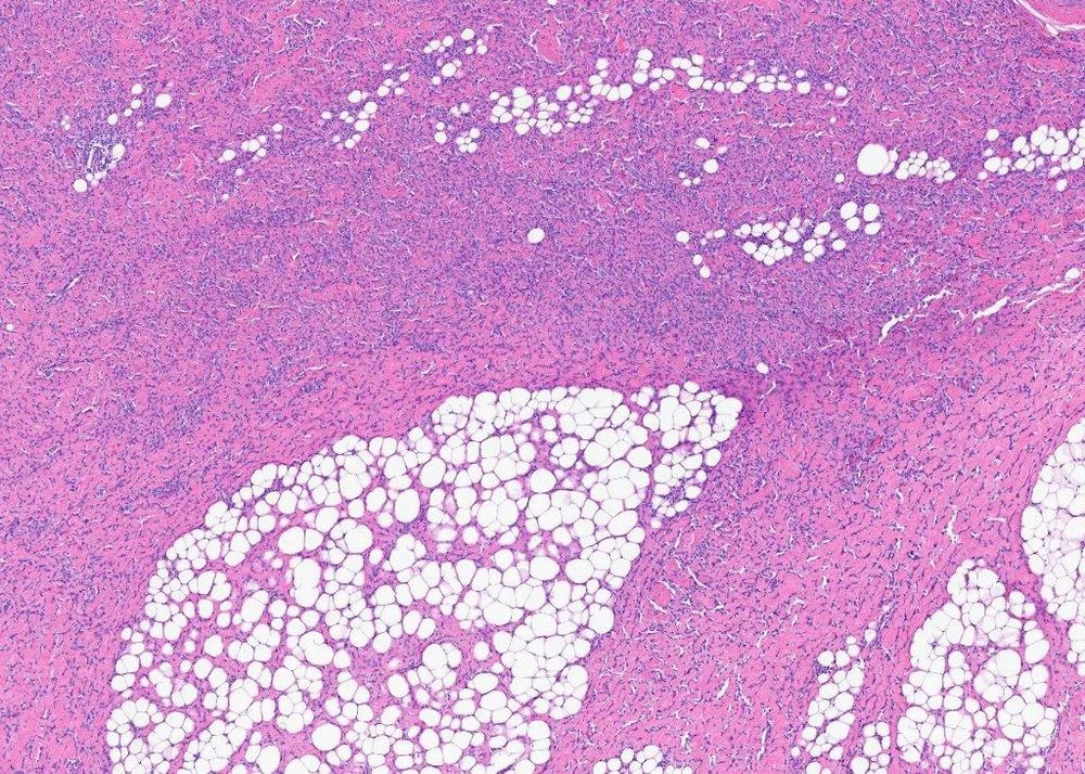
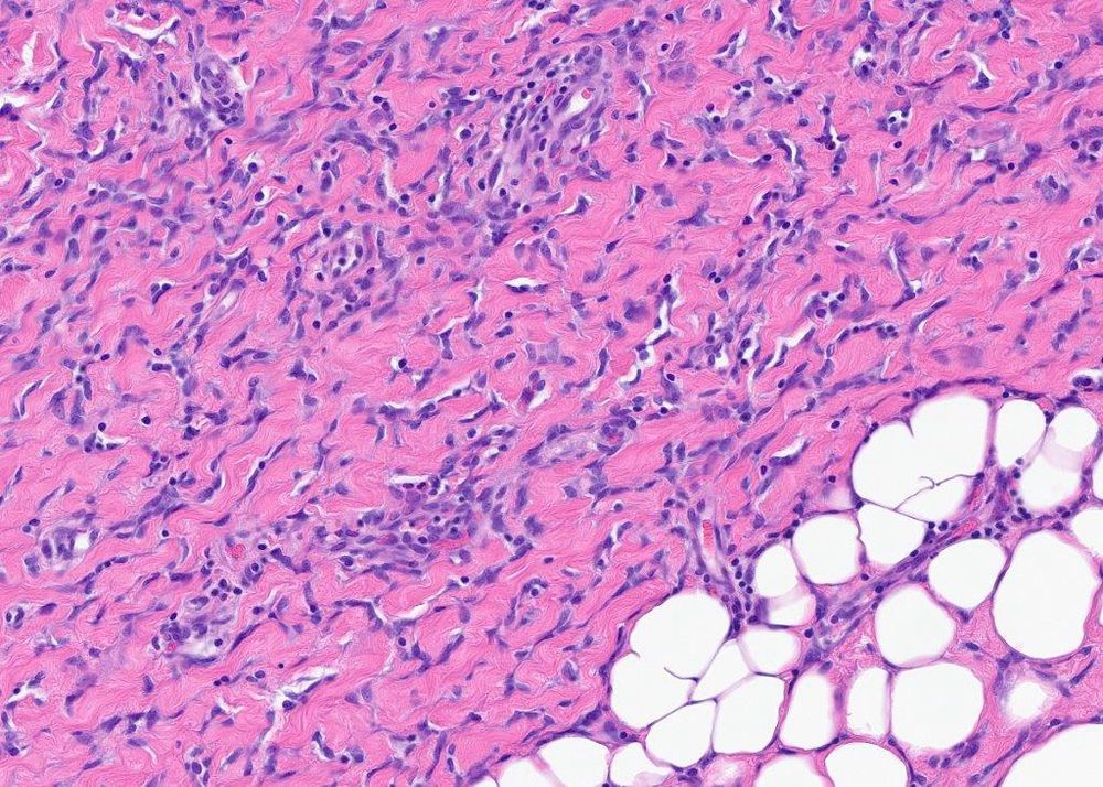
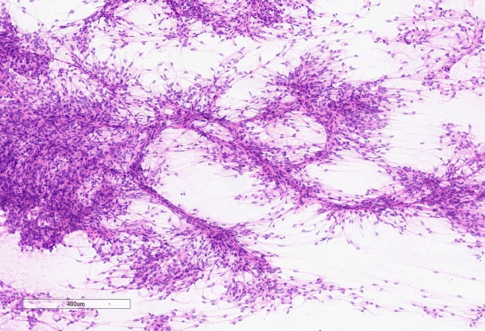
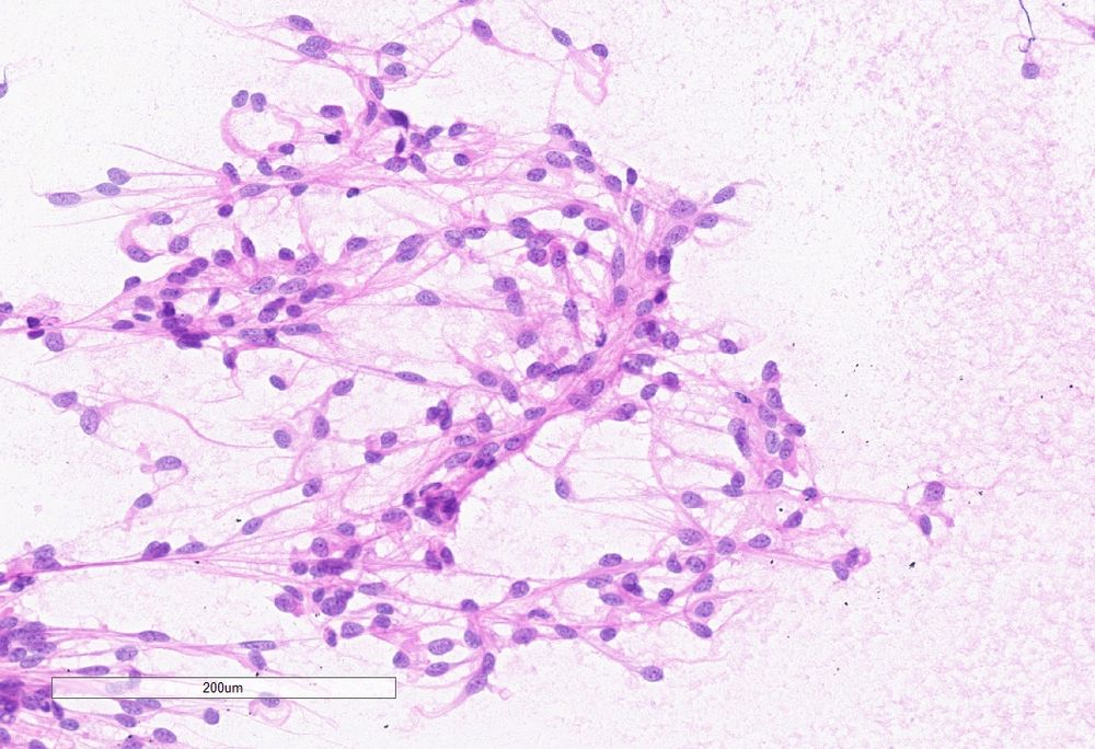
#PathSky I usually like to show cases where the morphology makes sense of the molecular, but here’s one where you kind of need the molecular. Axillary mass, 9 month male. Very hyalinized, infiltration of fat, cracking artifact. Strongly CD34-pos. Giant cell fibroblastoma?
February 24, 2025 at 8:32 PM
#PathSky I usually like to show cases where the morphology makes sense of the molecular, but here’s one where you kind of need the molecular. Axillary mass, 9 month male. Very hyalinized, infiltration of fat, cracking artifact. Strongly CD34-pos. Giant cell fibroblastoma?
Reposted by LDMMarcelis
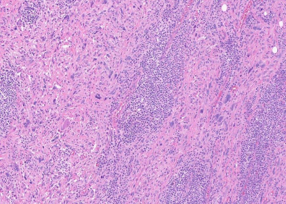
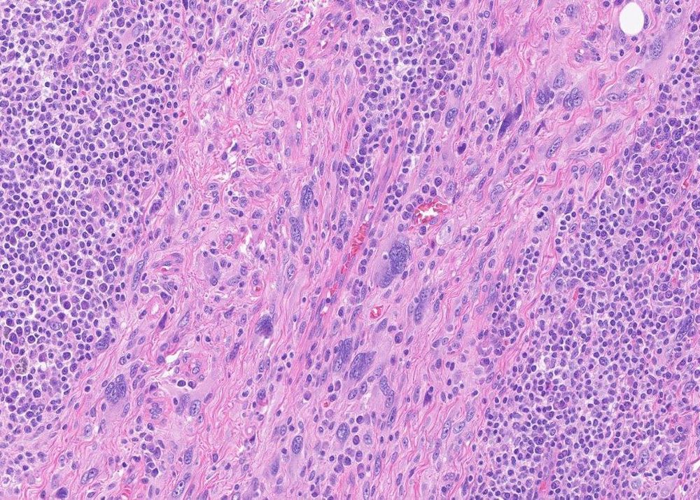
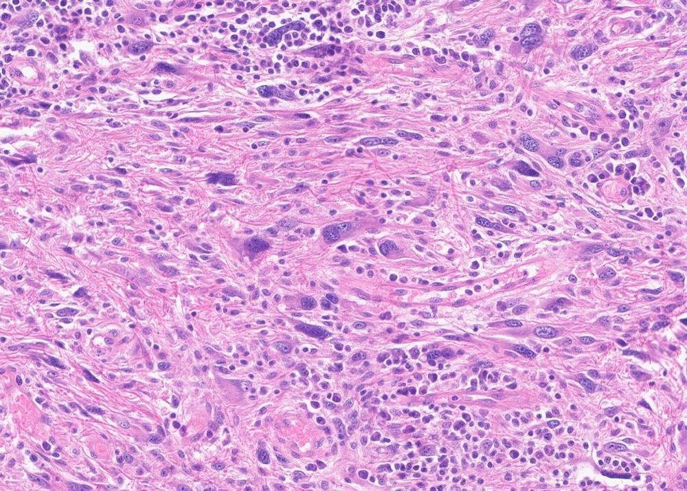
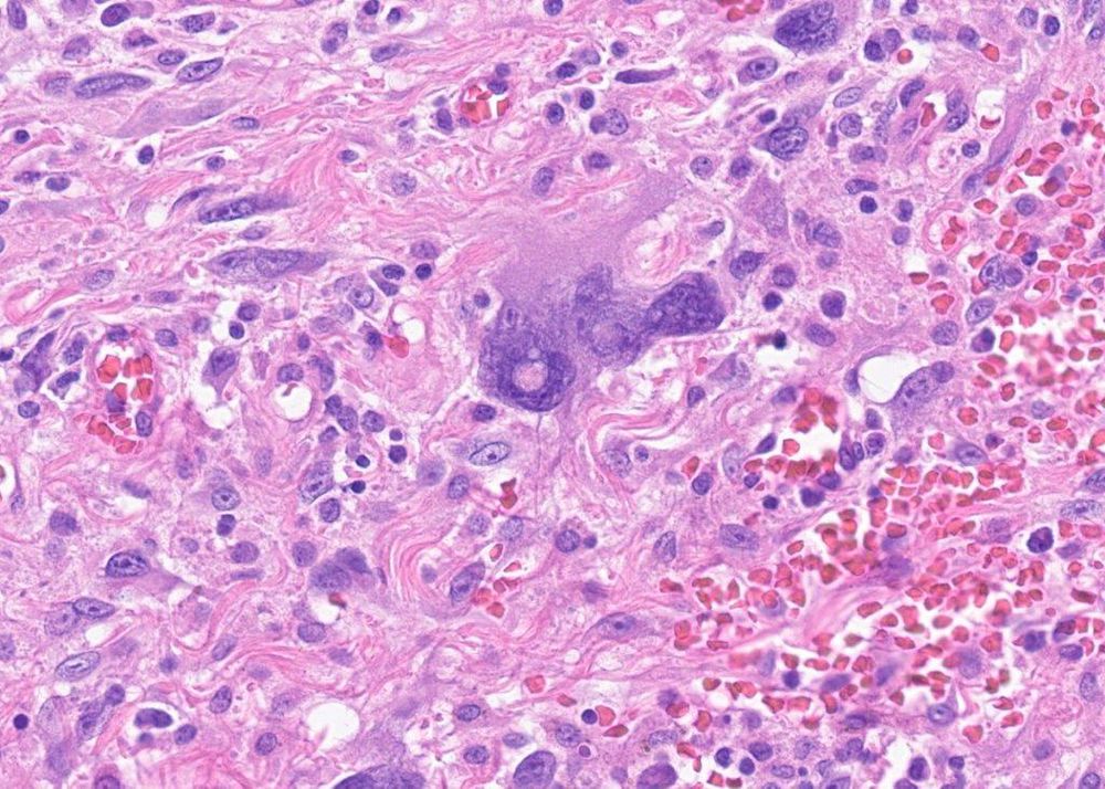
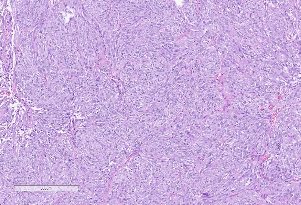
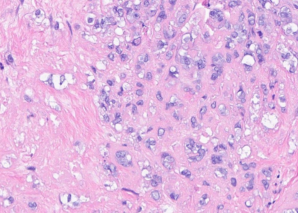
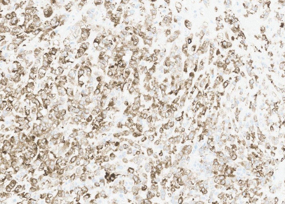
#PathSky 79M with a superficial neck mass. Wildly pleomorphic, quite cellular- undifferentiated pleomorphic sarcoma, right? There are basically no mitoses though, the cells have abundant granular-glassy cytoplasm, there is a lot of inflammation, and many bizarre cells have nuclear inclusions.
February 5, 2025 at 7:08 PM
#PathSky 79M with a superficial neck mass. Wildly pleomorphic, quite cellular- undifferentiated pleomorphic sarcoma, right? There are basically no mitoses though, the cells have abundant granular-glassy cytoplasm, there is a lot of inflammation, and many bizarre cells have nuclear inclusions.
Reposted by LDMMarcelis
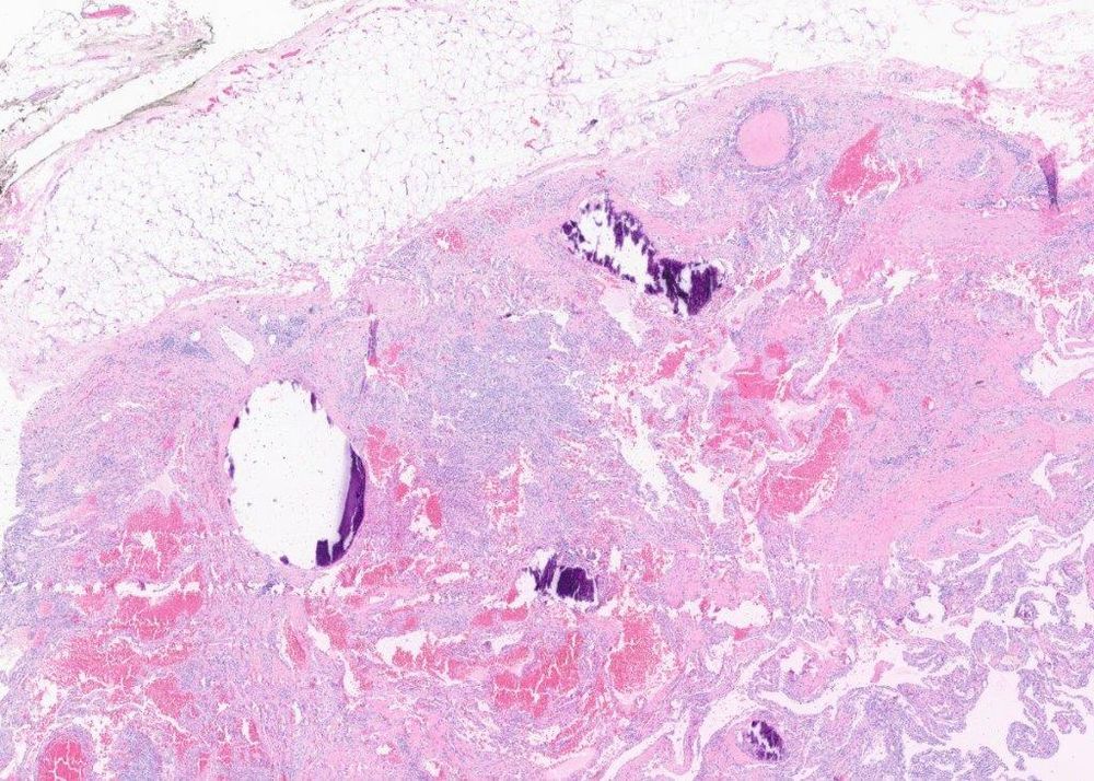
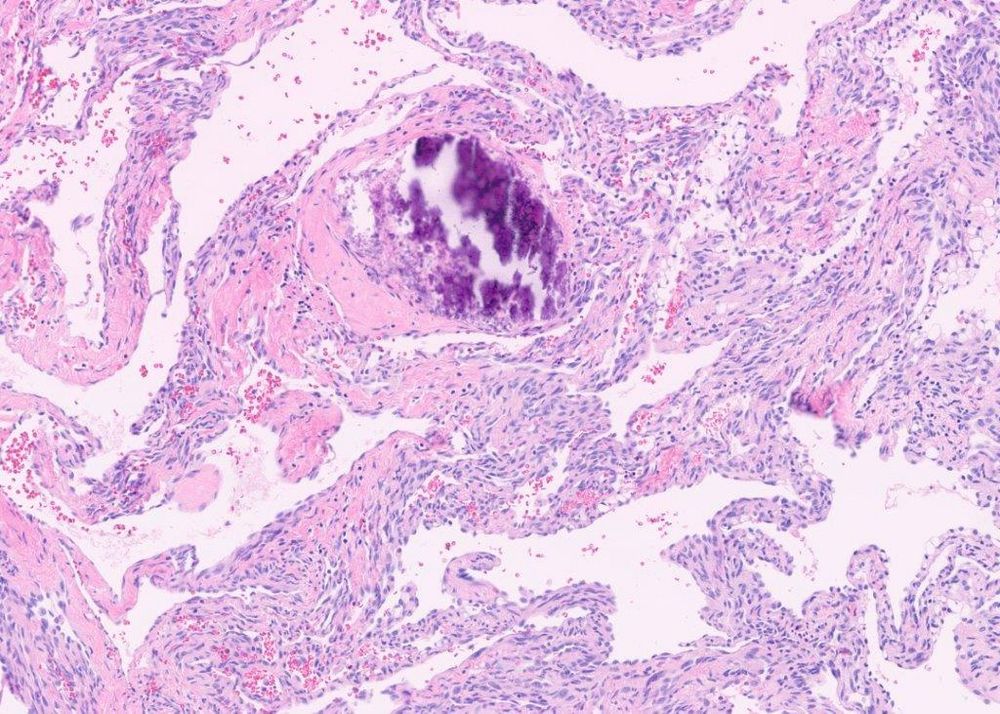
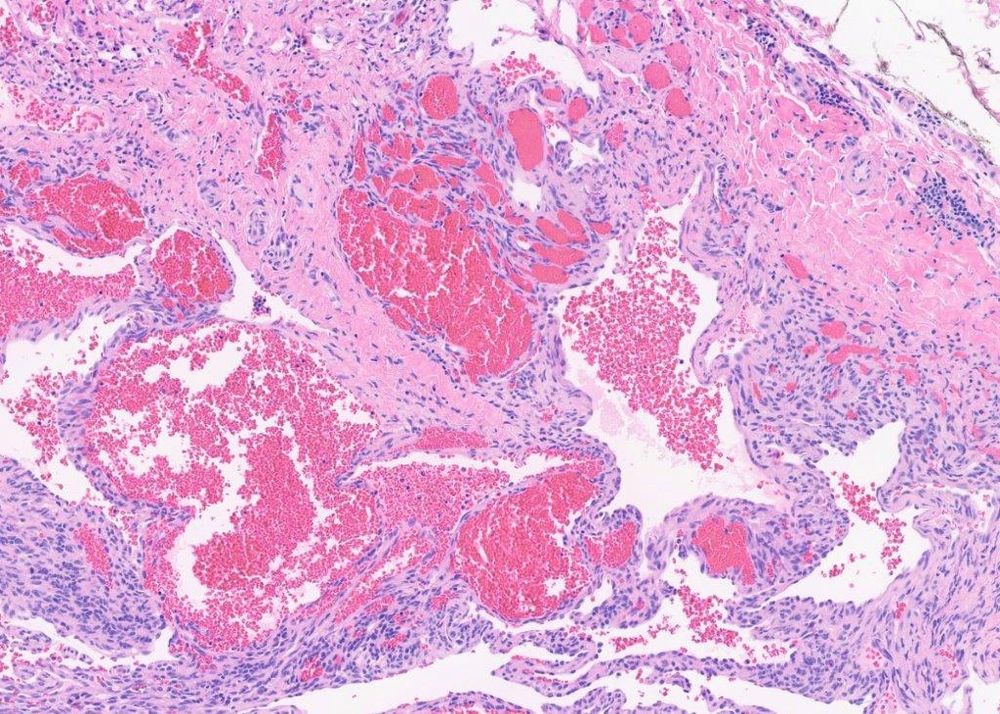
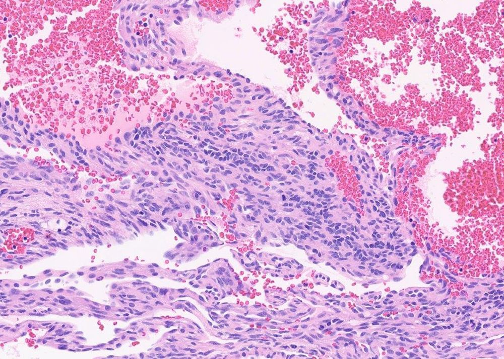
#PathSky Slowly growing superficial leg mass, 38M. Well-circumscribed, phleboliths, thrombosed vessels, cellular spindle cell areas, cavernous vascular channels. Dx?
February 5, 2025 at 6:57 PM
#PathSky Slowly growing superficial leg mass, 38M. Well-circumscribed, phleboliths, thrombosed vessels, cellular spindle cell areas, cavernous vascular channels. Dx?
Reposted by LDMMarcelis

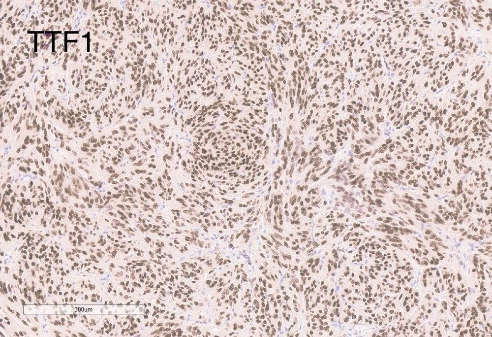
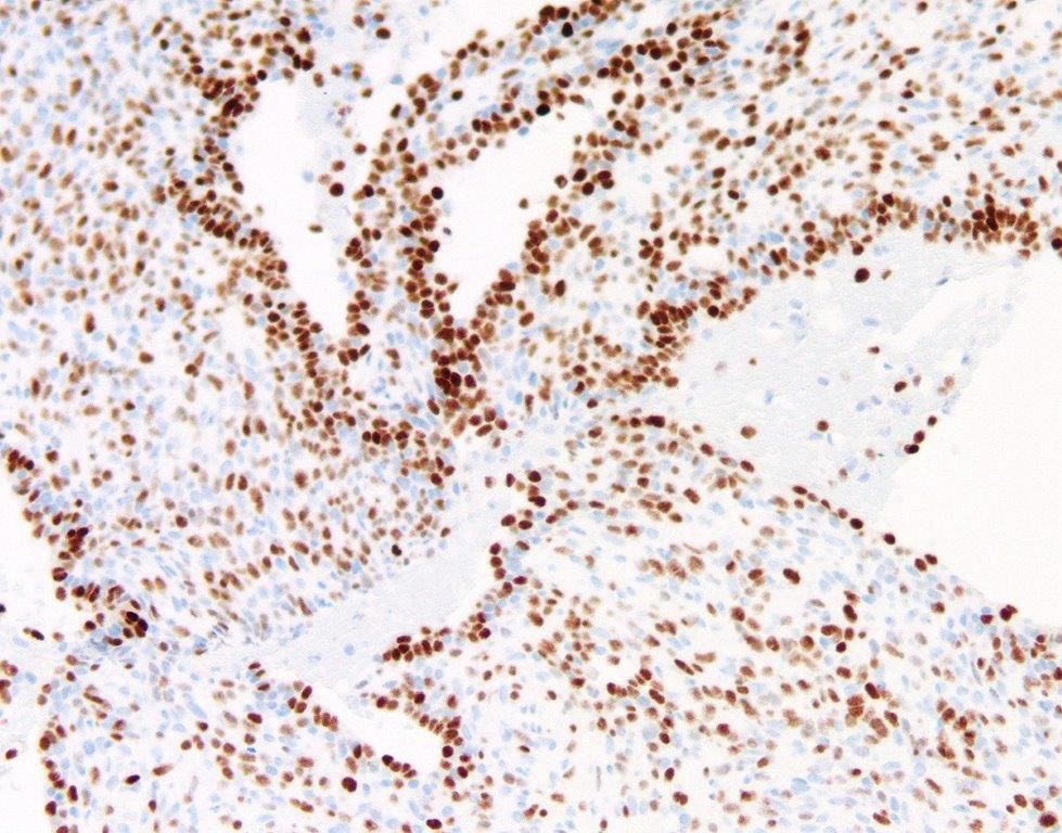
Just like Texas rarely sees snow ❄️, every once in a while you will find a primary tumor of the posterior pituitary so keep it in mind during frozen section analysis! Here is a classic example of pituicytoma showing diffuse nuclear positivity for TTF1 #pathsky #neuropath #medsky 🔬 🧠
January 19, 2025 at 8:58 PM
Just like Texas rarely sees snow ❄️, every once in a while you will find a primary tumor of the posterior pituitary so keep it in mind during frozen section analysis! Here is a classic example of pituicytoma showing diffuse nuclear positivity for TTF1 #pathsky #neuropath #medsky 🔬 🧠
Reposted by LDMMarcelis
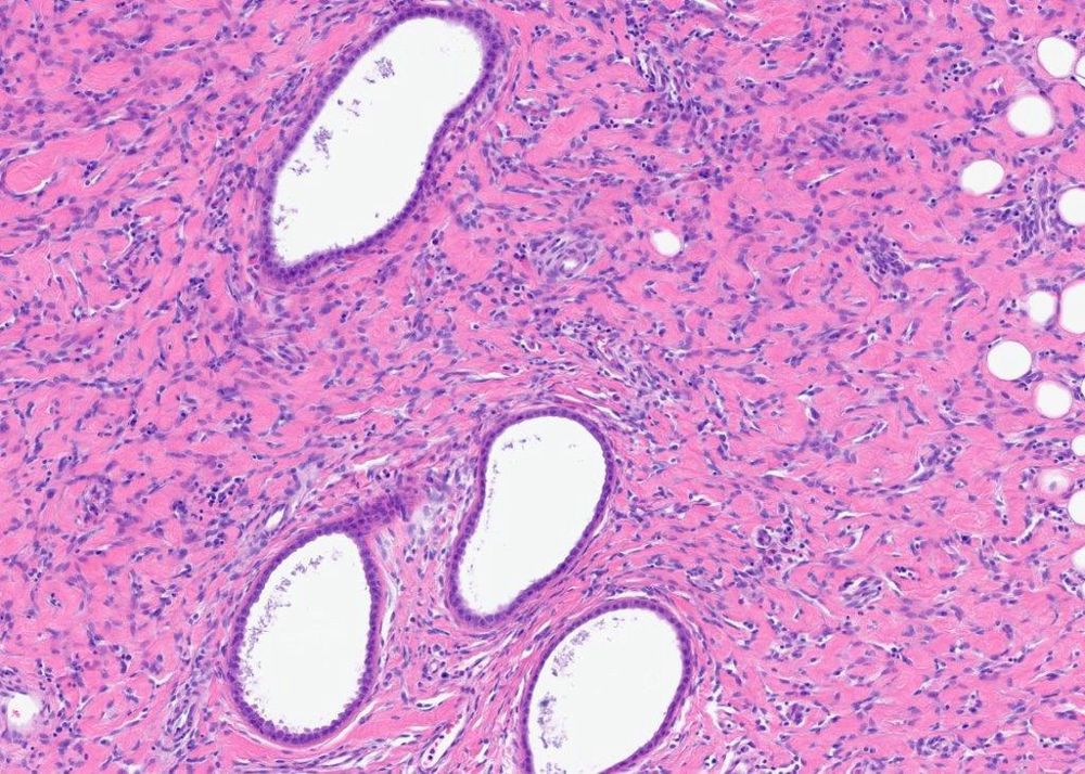
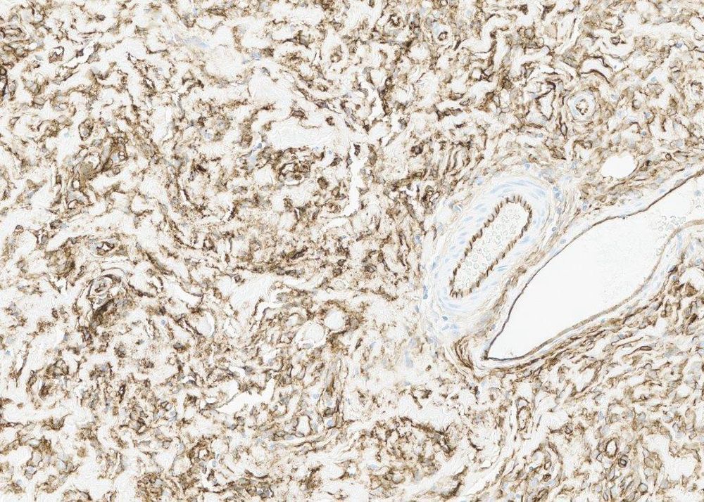
#PathSky 37M with a large retroperitoneal mass. Big, fatty thing. The outside quite rightly worried about well-differentiated LPS and a lipomatous angiomyolipoma, but MDM2, SMA and melanocytic markers were negative. Some WDL are MDM2-neg/MDM4-pos. Should we do more molecular ? Or something else?


January 13, 2025 at 3:02 PM
#PathSky 37M with a large retroperitoneal mass. Big, fatty thing. The outside quite rightly worried about well-differentiated LPS and a lipomatous angiomyolipoma, but MDM2, SMA and melanocytic markers were negative. Some WDL are MDM2-neg/MDM4-pos. Should we do more molecular ? Or something else?
Reposted by LDMMarcelis
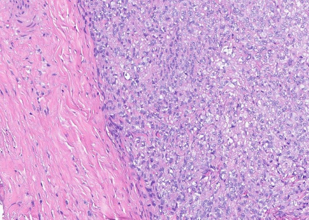
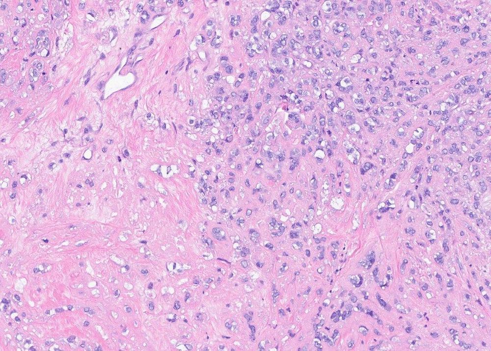
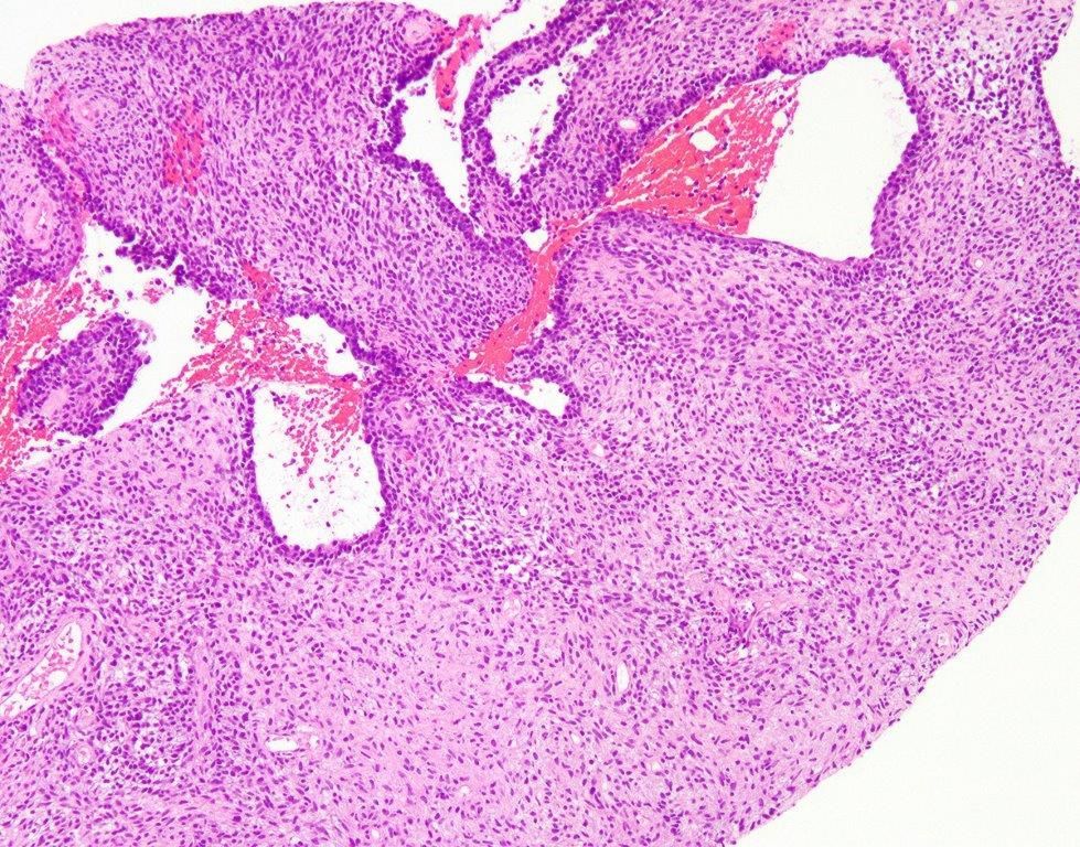
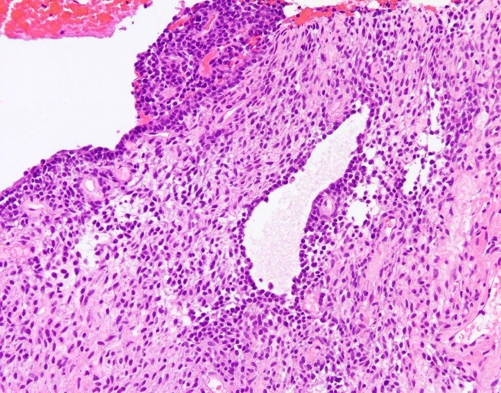
#PathSky 32M with extra-axial, dural-based, frontoparietal mass. Clearly malignant epithelioid cells arranged in cords and nests in a sclerotic background. Looks a lot like a sclerosing epithelioid fibrosarcoma, and it is strongly MUC4-positive. So, we’re done, right?
January 8, 2025 at 5:13 PM
#PathSky 32M with extra-axial, dural-based, frontoparietal mass. Clearly malignant epithelioid cells arranged in cords and nests in a sclerotic background. Looks a lot like a sclerosing epithelioid fibrosarcoma, and it is strongly MUC4-positive. So, we’re done, right?
Reposted by LDMMarcelis

January 3, 2025 at 2:19 PM
Reposted by LDMMarcelis

✨ Happy New Year! We're excited to connect with the pathology community here on Bluesky. 🏷️Tag your pathology friends to let them know we're here!
#Pathology #PathSky #PathologyOutlines
#Pathology #PathSky #PathologyOutlines

January 2, 2025 at 6:10 PM
✨ Happy New Year! We're excited to connect with the pathology community here on Bluesky. 🏷️Tag your pathology friends to let them know we're here!
#Pathology #PathSky #PathologyOutlines
#Pathology #PathSky #PathologyOutlines
Reposted by LDMMarcelis
#PathSky Dear Pathologists everywhere: Please, please, please. Do not entirely submit an 11cm skin excision entirely, in 164 blocks. Yours, Whoever it is You Send Your Consults to.
December 26, 2024 at 2:42 PM
#PathSky Dear Pathologists everywhere: Please, please, please. Do not entirely submit an 11cm skin excision entirely, in 164 blocks. Yours, Whoever it is You Send Your Consults to.
Reposted by LDMMarcelis
#PathSky 30M, abdominal wall mass. Microcysts, bland round to spindled cells, thick-walled vessels. Positive for desmin, MyoD1, myogenin, and GFAP. Classic findings in EWSR1::PATZ1 sarcoma. Why isn’t this a fusion-positive RMS? Excellent question…https://pubmed.ncbi.nlm.nih.gov/38043789/
December 6, 2024 at 5:24 PM
#PathSky 30M, abdominal wall mass. Microcysts, bland round to spindled cells, thick-walled vessels. Positive for desmin, MyoD1, myogenin, and GFAP. Classic findings in EWSR1::PATZ1 sarcoma. Why isn’t this a fusion-positive RMS? Excellent question…https://pubmed.ncbi.nlm.nih.gov/38043789/
Reposted by LDMMarcelis


Reposted by LDMMarcelis
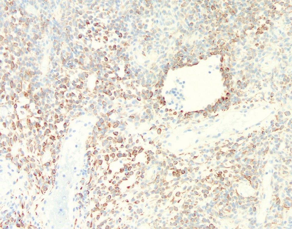
#PathSky Solid and cystic mass next to the liver in a 50F with no relevant hx or other masses. Once you think of it, extraspinal myxopapillary ependymoma in a rather odd location seems a good fit. Why not, right?
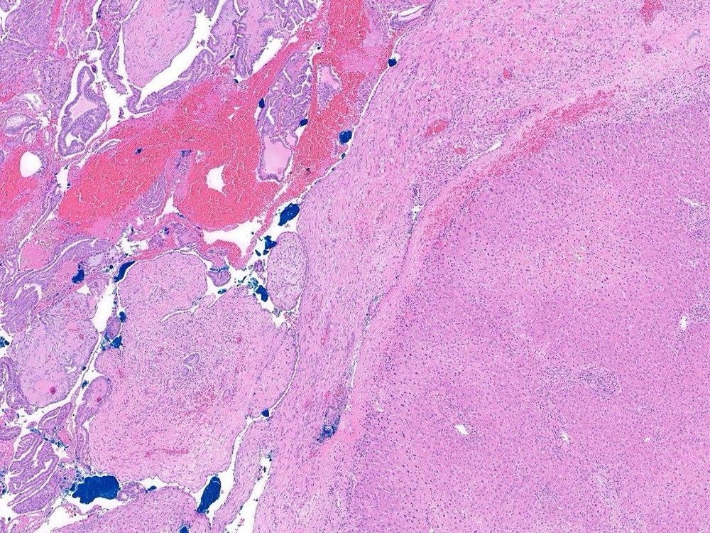
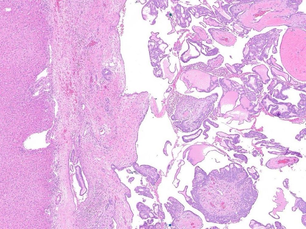
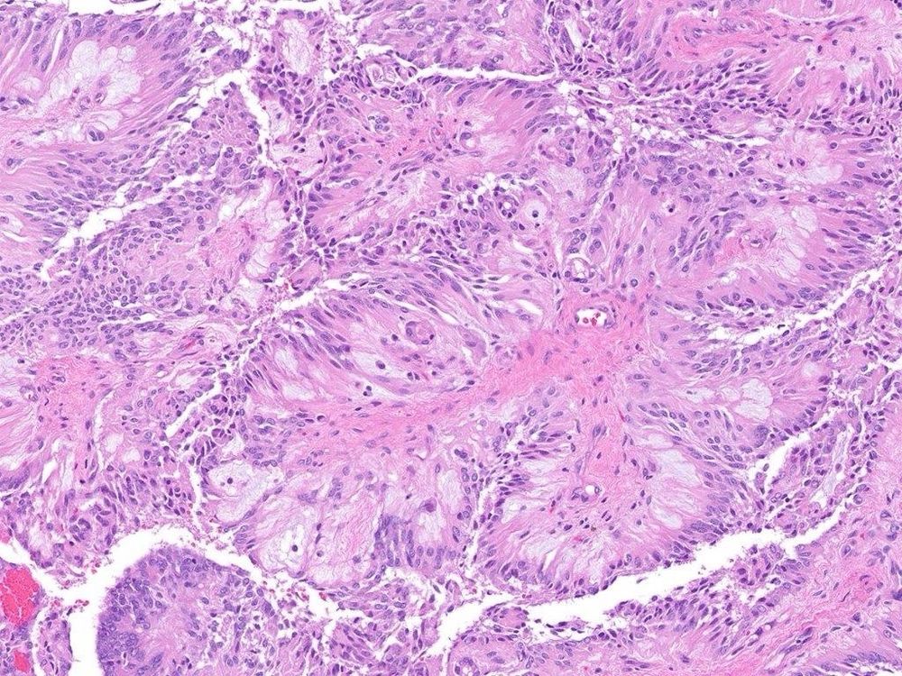
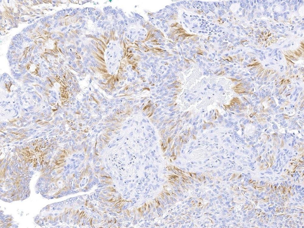
December 2, 2024 at 7:34 PM
#PathSky Solid and cystic mass next to the liver in a 50F with no relevant hx or other masses. Once you think of it, extraspinal myxopapillary ependymoma in a rather odd location seems a good fit. Why not, right?