


Kevin Staras
@kevinstaras.bsky.social
NeuroProf at University of Sussex, UK • Synapses, Circuits, Plasticity, Disease, Decision-making
https://www.thestaraslab.org/
https://www.thestaraslab.org/
Aw, we got the cover for our new paper on X-ray imaging and atlas building in the snail brain. www.pnas.org/doi/10.1073/... Thanks @pnas.org .. and to @sussexneuro.bsky.social @leverhulme.bsky.social @ukri.org for funding support #invertebrate #brain #neuroscience THREAD: bsky.app/profile/kevi...

March 5, 2025 at 10:24 AM
Aw, we got the cover for our new paper on X-ray imaging and atlas building in the snail brain. www.pnas.org/doi/10.1073/... Thanks @pnas.org .. and to @sussexneuro.bsky.social @leverhulme.bsky.social @ukri.org for funding support #invertebrate #brain #neuroscience THREAD: bsky.app/profile/kevi...
Michael Crossley led the experimental work, supported by Anna Simon, @arndroth.bsky.social and
@enzomarra.bsky.social Thanks to @sussexneuro.bsky.social @leverhulme.bsky.social @ukri.org and @diamondlightsource.bsky.social for funding support. Thanks for reading! 10/10
@enzomarra.bsky.social Thanks to @sussexneuro.bsky.social @leverhulme.bsky.social @ukri.org and @diamondlightsource.bsky.social for funding support. Thanks for reading! 10/10
February 28, 2025 at 9:40 AM
Michael Crossley led the experimental work, supported by Anna Simon, @arndroth.bsky.social and
@enzomarra.bsky.social Thanks to @sussexneuro.bsky.social @leverhulme.bsky.social @ukri.org and @diamondlightsource.bsky.social for funding support. Thanks for reading! 10/10
@enzomarra.bsky.social Thanks to @sussexneuro.bsky.social @leverhulme.bsky.social @ukri.org and @diamondlightsource.bsky.social for funding support. Thanks for reading! 10/10
Our approach should readily generalize to other model systems with comparable brain sizes (e.g. other molluscs, crustacea, annelids, insects). On its own, it won’t yield a full wiring diagram, but it does rapidly provide a detailed overview map for atlas building and comparative studies. 9/10
February 28, 2025 at 9:38 AM
Our approach should readily generalize to other model systems with comparable brain sizes (e.g. other molluscs, crustacea, annelids, insects). On its own, it won’t yield a full wiring diagram, but it does rapidly provide a detailed overview map for atlas building and comparative studies. 9/10
This provides the locations of principal feeding-circuit cell types, including motoneurons, CPG neurons and modulatory cells, alongside a detailed summary of their main functional properties. 8/10

February 28, 2025 at 9:37 AM
This provides the locations of principal feeding-circuit cell types, including motoneurons, CPG neurons and modulatory cells, alongside a detailed summary of their main functional properties. 8/10
We also brought together the anatomical mapping and functional information to establish the beginnings of a fully scalable functional cell atlas of the brain of Lymnaea stagnalis: sites.google.com/view/snailbr... 7/10
February 28, 2025 at 9:36 AM
We also brought together the anatomical mapping and functional information to establish the beginnings of a fully scalable functional cell atlas of the brain of Lymnaea stagnalis: sites.google.com/view/snailbr... 7/10
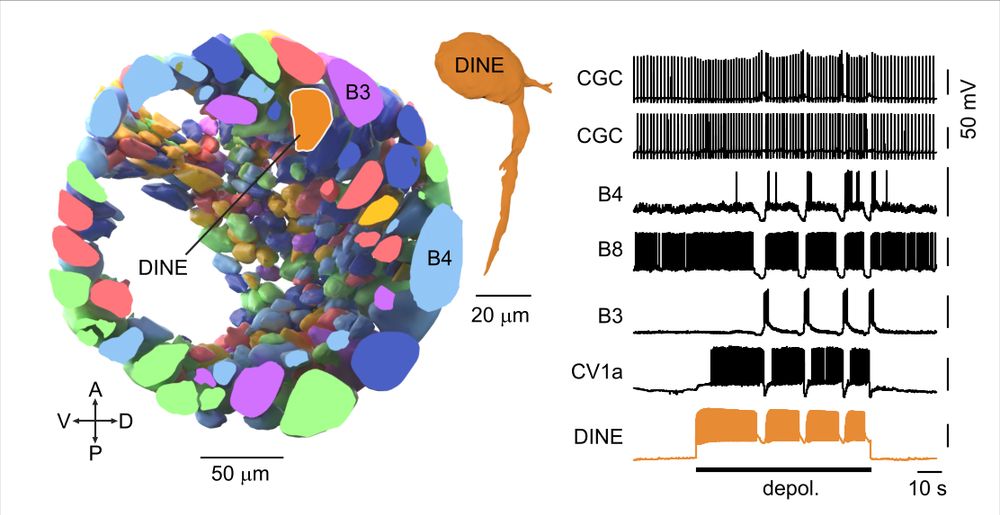
The consistent positioning of neurons across Lymnaea brains means the atlas can guide follow-up functional experiments. Targeting a non-superficial region led to the discovery of DINE (“Diamond Neuron”), an apt name 😜 because it activates the food ingestion circuitry. 6/10
February 28, 2025 at 9:35 AM
The consistent positioning of neurons across Lymnaea brains means the atlas can guide follow-up functional experiments. Targeting a non-superficial region led to the discovery of DINE (“Diamond Neuron”), an apt name 😜 because it activates the food ingestion circuitry. 6/10
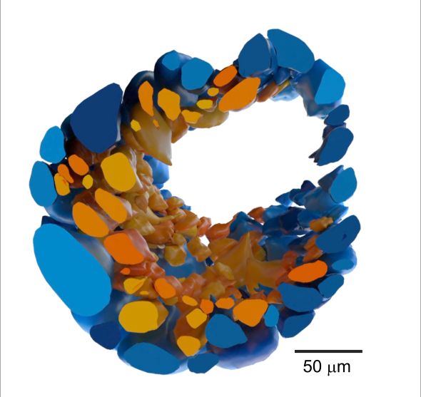
The 3D reconstruction revealed the organization of neurons beneath the surface layer for the first time. It turns out around half the neurons (coloured orange) are non-superficial - a hidden world of circuit components that can now be studied. 5/10
February 28, 2025 at 9:35 AM
The 3D reconstruction revealed the organization of neurons beneath the surface layer for the first time. It turns out around half the neurons (coloured orange) are non-superficial - a hidden world of circuit components that can now be studied. 5/10
We then used the excellent volume image-sharing, annotation, and reconstruction platform
@webknossos.org to fully reconstruct the buccal ganglia (one side is shown here) housing the main feeding circuitry, yielding the first accurate estimate of the total number of neurons: ~1100. 4/10
@webknossos.org to fully reconstruct the buccal ganglia (one side is shown here) housing the main feeding circuitry, yielding the first accurate estimate of the total number of neurons: ~1100. 4/10
February 28, 2025 at 9:34 AM
We then used the excellent volume image-sharing, annotation, and reconstruction platform
@webknossos.org to fully reconstruct the buccal ganglia (one side is shown here) housing the main feeding circuitry, yielding the first accurate estimate of the total number of neurons: ~1100. 4/10
@webknossos.org to fully reconstruct the buccal ganglia (one side is shown here) housing the main feeding circuitry, yielding the first accurate estimate of the total number of neurons: ~1100. 4/10
Michael Crossley led the experimental work, supported by Anna Simon, @arndroth.bsky.social and @enzomarra.bsky.social Thanks to @sussexneuro.bsky.social @leverhulme.bsky.social @ukri.org and @diamondlightsource.bsky.social for funding support. Thanks for reading! 10/10
February 28, 2025 at 8:49 AM
Michael Crossley led the experimental work, supported by Anna Simon, @arndroth.bsky.social and @enzomarra.bsky.social Thanks to @sussexneuro.bsky.social @leverhulme.bsky.social @ukri.org and @diamondlightsource.bsky.social for funding support. Thanks for reading! 10/10
Our approach should readily generalize to other model systems with comparable brain sizes (e.g. other molluscs, crustacea, annelids, insects). On its own, it won’t yield a full wiring diagram, but it does rapidly provide a detailed overview map for atlas building and comparative studies. 9/10
February 28, 2025 at 8:47 AM
Our approach should readily generalize to other model systems with comparable brain sizes (e.g. other molluscs, crustacea, annelids, insects). On its own, it won’t yield a full wiring diagram, but it does rapidly provide a detailed overview map for atlas building and comparative studies. 9/10
This provides the locations of principal feeding-circuit cell types, including motoneurons, CPG neurons and modulatory cells, alongside a detailed summary of their main functional properties. 8/10
February 28, 2025 at 8:45 AM
This provides the locations of principal feeding-circuit cell types, including motoneurons, CPG neurons and modulatory cells, alongside a detailed summary of their main functional properties. 8/10
We also brought together the anatomical mapping and functional information to establish the beginnings of a fully scalable functional cell atlas of the brain of Lymnaea stagnalis: sites.google.com/view/snailbr... 7/10
February 28, 2025 at 8:44 AM
We also brought together the anatomical mapping and functional information to establish the beginnings of a fully scalable functional cell atlas of the brain of Lymnaea stagnalis: sites.google.com/view/snailbr... 7/10
The consistent positioning of neurons across Lymnaea brains means the atlas can guide follow-up functional experiments. Targeting a non-superficial region led to the discovery of DINE (“Diamond Neuron”), an apt name 😜 because it activates the food ingestion circuitry. 6/10
February 28, 2025 at 8:44 AM
The consistent positioning of neurons across Lymnaea brains means the atlas can guide follow-up functional experiments. Targeting a non-superficial region led to the discovery of DINE (“Diamond Neuron”), an apt name 😜 because it activates the food ingestion circuitry. 6/10
The 3D reconstruction revealed the organization of neurons beneath the surface layer for the first time. It turns out around half the neurons (coloured orange) are non-superficial - a hidden world of circuit components that can now be studied. 5/10
February 28, 2025 at 8:42 AM
The 3D reconstruction revealed the organization of neurons beneath the surface layer for the first time. It turns out around half the neurons (coloured orange) are non-superficial - a hidden world of circuit components that can now be studied. 5/10
We then used the excellent volume image-sharing, annotation, and reconstruction platform @webknossos.org to fully reconstruct the buccal ganglia (one side is shown here) housing the main feeding circuitry, yielding the first accurate estimate of the total number of neurons: ~1100. 4/10
February 28, 2025 at 8:41 AM
We then used the excellent volume image-sharing, annotation, and reconstruction platform @webknossos.org to fully reconstruct the buccal ganglia (one side is shown here) housing the main feeding circuitry, yielding the first accurate estimate of the total number of neurons: ~1100. 4/10
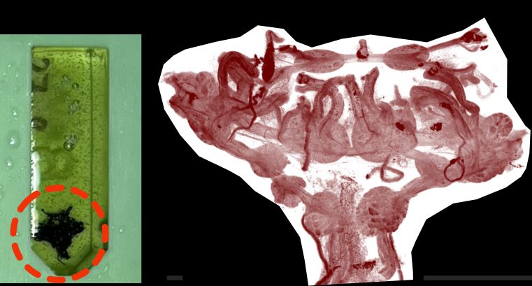
We plastic-embedded the 3x3x2 mm3 brain and performed X-ray tomography imaging at the Diamond Synchrotron Facility @diamondlightsource.bsky.social
February 28, 2025 at 8:38 AM
We plastic-embedded the 3x3x2 mm3 brain and performed X-ray tomography imaging at the Diamond Synchrotron Facility @diamondlightsource.bsky.social
The mollusc Lymnaea is a classical system for neural circuit studies. However, we lack a cell-level atlas of its multi-mm scale brain to guide functional investigations. The solution? 2/10
February 28, 2025 at 8:37 AM
The mollusc Lymnaea is a classical system for neural circuit studies. However, we lack a cell-level atlas of its multi-mm scale brain to guide functional investigations. The solution? 2/10
Looking for a quick way to map the brain of your favourite small animal model?
See our new paper. www.pnas.org/doi/10.1073/... 1/10
See our new paper. www.pnas.org/doi/10.1073/... 1/10
February 28, 2025 at 8:36 AM
Looking for a quick way to map the brain of your favourite small animal model?
See our new paper. www.pnas.org/doi/10.1073/... 1/10
See our new paper. www.pnas.org/doi/10.1073/... 1/10
Mix neuroscience with the seaside! 4 year PhD programme on the UK's south coast at the University of Sussex. Fantastic community of excellent researchers across the neuro field. Apply now. Please RT.

November 27, 2024 at 1:11 PM
Mix neuroscience with the seaside! 4 year PhD programme on the UK's south coast at the University of Sussex. Fantastic community of excellent researchers across the neuro field. Apply now. Please RT.