


Julia Eckert
@juliaeckert.bsky.social
@dfg.de Walter Benjamin Fellow & Postdoctoral Researcher @yap-lab.bsky.social | Co-organizer @epimechfc.bsky.social | Mechanobiology - Cell & Tissue Mechanics - Soft Matter Physics - Image Analysis
👉 https://julia-eckert.github.io
👉 https://julia-eckert.github.io
Excited to participate in the Scientific Art Contest at #CellMech2025.

October 2, 2025 at 1:55 PM
Excited to participate in the Scientific Art Contest at #CellMech2025.
The last paper from my PhD is now online! 🥳
👉 pubs.aip.org/aip/rsi/arti...
We have designed a new device based on the elastic micropillar array assay to measure the detachment forces between spread cells.
@leidenphysics.bsky.social
👉 pubs.aip.org/aip/rsi/arti...
We have designed a new device based on the elastic micropillar array assay to measure the detachment forces between spread cells.
@leidenphysics.bsky.social

September 5, 2025 at 1:11 PM
The last paper from my PhD is now online! 🥳
👉 pubs.aip.org/aip/rsi/arti...
We have designed a new device based on the elastic micropillar array assay to measure the detachment forces between spread cells.
@leidenphysics.bsky.social
👉 pubs.aip.org/aip/rsi/arti...
We have designed a new device based on the elastic micropillar array assay to measure the detachment forces between spread cells.
@leidenphysics.bsky.social
Now published in @natcomms.nature.com! 🥳
👉 rdcu.be/eATn3
We developed image analysis tools to capture the nematic orientation field of 3D tissue surfaces. Tested on epithelial aggregates, zebrafish hearts, myoblasts on spheres & micro-vessels, we combined soft matter physics with exp. biology.
👉 rdcu.be/eATn3
We developed image analysis tools to capture the nematic orientation field of 3D tissue surfaces. Tested on epithelial aggregates, zebrafish hearts, myoblasts on spheres & micro-vessels, we combined soft matter physics with exp. biology.

August 15, 2025 at 12:13 PM
Now published in @natcomms.nature.com! 🥳
👉 rdcu.be/eATn3
We developed image analysis tools to capture the nematic orientation field of 3D tissue surfaces. Tested on epithelial aggregates, zebrafish hearts, myoblasts on spheres & micro-vessels, we combined soft matter physics with exp. biology.
👉 rdcu.be/eATn3
We developed image analysis tools to capture the nematic orientation field of 3D tissue surfaces. Tested on epithelial aggregates, zebrafish hearts, myoblasts on spheres & micro-vessels, we combined soft matter physics with exp. biology.
A fantastic week at the Mechanobiology Conference in Vietnam, filled with fascinating talks, fruitful discussions, and amazing people.
Huge thanks to the organizers and ICISE for making it all possible!
Huge thanks to the organizers and ICISE for making it all possible!

July 11, 2025 at 4:49 PM
A fantastic week at the Mechanobiology Conference in Vietnam, filled with fascinating talks, fruitful discussions, and amazing people.
Huge thanks to the organizers and ICISE for making it all possible!
Huge thanks to the organizers and ICISE for making it all possible!
Please check out this fantastic work by my friend Svetlana Jovanic 👇
'Resilience in zebrafish embryoids'
www.biorxiv.org/content/10.1...
'Resilience in zebrafish embryoids'
www.biorxiv.org/content/10.1...
June 20, 2025 at 10:27 PM
Please check out this fantastic work by my friend Svetlana Jovanic 👇
'Resilience in zebrafish embryoids'
www.biorxiv.org/content/10.1...
'Resilience in zebrafish embryoids'
www.biorxiv.org/content/10.1...
Our new review paper from @yap-lab.bsky.social is online - together with Virgile Viasnoff!
👉 'New directions in epithelial mechanoadaptation'
www.sciencedirect.com/science/arti...
👉 'New directions in epithelial mechanoadaptation'
www.sciencedirect.com/science/arti...

May 25, 2025 at 2:42 AM
Our new review paper from @yap-lab.bsky.social is online - together with Virgile Viasnoff!
👉 'New directions in epithelial mechanoadaptation'
www.sciencedirect.com/science/arti...
👉 'New directions in epithelial mechanoadaptation'
www.sciencedirect.com/science/arti...
Started my first day as a Walter Benjamin Fellow! 🥳
Looking forward to study symmetry-breaking in morphogenesis using and connecting concepts from soft matter physics & cell/molecular biology @yap-lab.bsky.social. 👉 physics meets biology!
Big thanks to the @dfg.de for funding my research.
Looking forward to study symmetry-breaking in morphogenesis using and connecting concepts from soft matter physics & cell/molecular biology @yap-lab.bsky.social. 👉 physics meets biology!
Big thanks to the @dfg.de for funding my research.

May 1, 2025 at 3:27 AM
Started my first day as a Walter Benjamin Fellow! 🥳
Looking forward to study symmetry-breaking in morphogenesis using and connecting concepts from soft matter physics & cell/molecular biology @yap-lab.bsky.social. 👉 physics meets biology!
Big thanks to the @dfg.de for funding my research.
Looking forward to study symmetry-breaking in morphogenesis using and connecting concepts from soft matter physics & cell/molecular biology @yap-lab.bsky.social. 👉 physics meets biology!
Big thanks to the @dfg.de for funding my research.
Congratulations to Denni from our @yap-lab.bsky.social team and all authors on the new paper! 🥳
'Advecting scaffolds: Controlling the remodeling of actomyosin with anillin' 👇
journals.aps.org/pre/abstract...
'Advecting scaffolds: Controlling the remodeling of actomyosin with anillin' 👇
journals.aps.org/pre/abstract...

February 10, 2025 at 8:18 AM
Congratulations to Denni from our @yap-lab.bsky.social team and all authors on the new paper! 🥳
'Advecting scaffolds: Controlling the remodeling of actomyosin with anillin' 👇
journals.aps.org/pre/abstract...
'Advecting scaffolds: Controlling the remodeling of actomyosin with anillin' 👇
journals.aps.org/pre/abstract...
… F-actin signal anti-correlates with the mean curvature, H, in Region I & II for each developmental stage. Assuming F-actin represents surface tension, gamma, the results correlate with Laplace’s law (P ~ gamma*H) for constant pressure!
👉 Linking biological properties with physical interpretations
👉 Linking biological properties with physical interpretations

January 29, 2025 at 11:01 PM
… F-actin signal anti-correlates with the mean curvature, H, in Region I & II for each developmental stage. Assuming F-actin represents surface tension, gamma, the results correlate with Laplace’s law (P ~ gamma*H) for constant pressure!
👉 Linking biological properties with physical interpretations
👉 Linking biological properties with physical interpretations
The sarcomeric network composed of acto-myosin contracts the heart … it pumps. We used the same tangent planes and analyzed the fluorescent signal of F-actin …
(Directors: high intensity = red, low = blue)
👉 Spatio-temporal correlations
(Directors: high intensity = red, low = blue)
👉 Spatio-temporal correlations

January 29, 2025 at 11:01 PM
The sarcomeric network composed of acto-myosin contracts the heart … it pumps. We used the same tangent planes and analyzed the fluorescent signal of F-actin …
(Directors: high intensity = red, low = blue)
👉 Spatio-temporal correlations
(Directors: high intensity = red, low = blue)
👉 Spatio-temporal correlations
By selecting data points, we can compare properties such as alignment of cells, i.e. local nematic order, between different regions (Region I: outer curvature, Region II: axes) in the heart & between different developmental stages (72, 120 hpf).
Region I: high alignment -> in direction of tension?
Region I: high alignment -> in direction of tension?

January 29, 2025 at 11:01 PM
By selecting data points, we can compare properties such as alignment of cells, i.e. local nematic order, between different regions (Region I: outer curvature, Region II: axes) in the heart & between different developmental stages (72, 120 hpf).
Region I: high alignment -> in direction of tension?
Region I: high alignment -> in direction of tension?
Spheroids are simple systems with only convex curvature. What about more complex systems?
We teamed up with @tobyandrews.bsky.social & @rashmi-priya.bsky.social, and analyzed the ventricular myocardium of Zebrafish hearts ... and it works! 👇
(Directors: high alignment = red, misaligned = blue)
We teamed up with @tobyandrews.bsky.social & @rashmi-priya.bsky.social, and analyzed the ventricular myocardium of Zebrafish hearts ... and it works! 👇
(Directors: high alignment = red, misaligned = blue)
January 29, 2025 at 11:01 PM
Spheroids are simple systems with only convex curvature. What about more complex systems?
We teamed up with @tobyandrews.bsky.social & @rashmi-priya.bsky.social, and analyzed the ventricular myocardium of Zebrafish hearts ... and it works! 👇
(Directors: high alignment = red, misaligned = blue)
We teamed up with @tobyandrews.bsky.social & @rashmi-priya.bsky.social, and analyzed the ventricular myocardium of Zebrafish hearts ... and it works! 👇
(Directors: high alignment = red, misaligned = blue)
As expected, our spherical aggregates have a total topological charge of +2.
We show that the number of defects correlates with the surface area, as previously reported in the theoretical literature, for example by Henkes et al, 2020.
👉 Experiment verifies theory
We show that the number of defects correlates with the surface area, as previously reported in the theoretical literature, for example by Henkes et al, 2020.
👉 Experiment verifies theory

January 29, 2025 at 11:01 PM
As expected, our spherical aggregates have a total topological charge of +2.
We show that the number of defects correlates with the surface area, as previously reported in the theoretical literature, for example by Henkes et al, 2020.
👉 Experiment verifies theory
We show that the number of defects correlates with the surface area, as previously reported in the theoretical literature, for example by Henkes et al, 2020.
👉 Experiment verifies theory
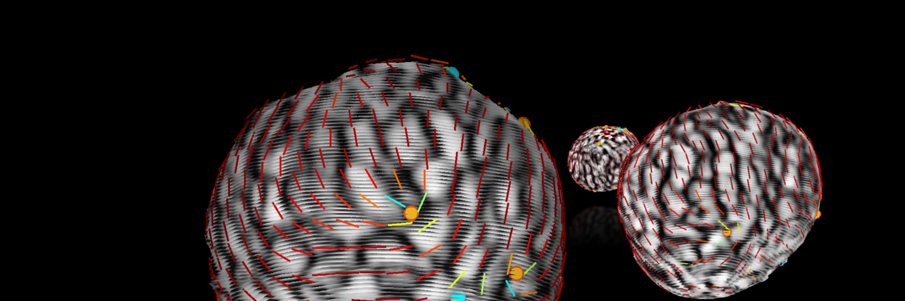
Nematic directors (white rods) align with cell orientation & spheroid surface, highlighting a big difference compared to 2D intensity projections of multiple image planes.
Our method allows not only the generation of the orientation field, but also the calculation of nematic topological defects.
Our method allows not only the generation of the orientation field, but also the calculation of nematic topological defects.

January 29, 2025 at 11:01 PM
Nematic directors (white rods) align with cell orientation & spheroid surface, highlighting a big difference compared to 2D intensity projections of multiple image planes.
Our method allows not only the generation of the orientation field, but also the calculation of nematic topological defects.
Our method allows not only the generation of the orientation field, but also the calculation of nematic topological defects.
Yes, but how?
We have developed an analysis pipeline that slices the outer layer of the cell system via tangent planes and analyzes the properties of these 2D-generated images. The center information, where the region is approximately flat, is then transferred back to the 3D surface.
Input: z-stack
We have developed an analysis pipeline that slices the outer layer of the cell system via tangent planes and analyzes the properties of these 2D-generated images. The center information, where the region is approximately flat, is then transferred back to the 3D surface.
Input: z-stack

January 29, 2025 at 11:01 PM
Yes, but how?
We have developed an analysis pipeline that slices the outer layer of the cell system via tangent planes and analyzes the properties of these 2D-generated images. The center information, where the region is approximately flat, is then transferred back to the 3D surface.
Input: z-stack
We have developed an analysis pipeline that slices the outer layer of the cell system via tangent planes and analyzes the properties of these 2D-generated images. The center information, where the region is approximately flat, is then transferred back to the 3D surface.
Input: z-stack
Thrilled to share our new preprint on detecting the nematic orientation field on surfaces of 3D multicellular systems!
www.biorxiv.org/content/10.1...
Huge thanks to all authors: @tobyandrews.bsky.social,Joe Pollard, @rashmi-priya.bsky.social, @yap-lab.bsky.social &Richard Morris
Any 3D systems?👇
www.biorxiv.org/content/10.1...
Huge thanks to all authors: @tobyandrews.bsky.social,Joe Pollard, @rashmi-priya.bsky.social, @yap-lab.bsky.social &Richard Morris
Any 3D systems?👇
January 29, 2025 at 11:01 PM
Thrilled to share our new preprint on detecting the nematic orientation field on surfaces of 3D multicellular systems!
www.biorxiv.org/content/10.1...
Huge thanks to all authors: @tobyandrews.bsky.social,Joe Pollard, @rashmi-priya.bsky.social, @yap-lab.bsky.social &Richard Morris
Any 3D systems?👇
www.biorxiv.org/content/10.1...
Huge thanks to all authors: @tobyandrews.bsky.social,Joe Pollard, @rashmi-priya.bsky.social, @yap-lab.bsky.social &Richard Morris
Any 3D systems?👇
Nematic directors (white rods) align with cell orientation & spheroid surface, highlighting a big difference compared to 2D intensity projections of multiple image planes.
Our method allows not only the generation of the orientation field, but also the calculation of nematic topological defects.
Our method allows not only the generation of the orientation field, but also the calculation of nematic topological defects.
January 29, 2025 at 10:52 PM
Nematic directors (white rods) align with cell orientation & spheroid surface, highlighting a big difference compared to 2D intensity projections of multiple image planes.
Our method allows not only the generation of the orientation field, but also the calculation of nematic topological defects.
Our method allows not only the generation of the orientation field, but also the calculation of nematic topological defects.
Interesting findings by Jacob Notbohm's group. Nematic defects may form as a result of spatially coordinated patterns in cell forces and motion.
👉 arxiv.org/abs/2501.04827
👉 arxiv.org/abs/2501.04827

January 13, 2025 at 9:06 AM
Interesting findings by Jacob Notbohm's group. Nematic defects may form as a result of spatially coordinated patterns in cell forces and motion.
👉 arxiv.org/abs/2501.04827
👉 arxiv.org/abs/2501.04827
The crossover length scale, in which the hexatic and nematic order are equally present, depends on cell-cell adhesion and correlates with the monolayer density.
www.nature.com/articles/s41...
www.nature.com/articles/s41...

November 16, 2024 at 11:49 PM
The crossover length scale, in which the hexatic and nematic order are equally present, depends on cell-cell adhesion and correlates with the monolayer density.
www.nature.com/articles/s41...
www.nature.com/articles/s41...
However, nematic symmetry requires elongated cells. Are they elongated? Not always, and most cells are hexagons, indicating hexatic (p=6) symmetry.
Indeed, hexatic symmetry is dominant at small (single cell) and nematic at large (tissue) length scales.
www.nature.com/articles/s41...
Indeed, hexatic symmetry is dominant at small (single cell) and nematic at large (tissue) length scales.
www.nature.com/articles/s41...

November 16, 2024 at 11:49 PM
However, nematic symmetry requires elongated cells. Are they elongated? Not always, and most cells are hexagons, indicating hexatic (p=6) symmetry.
Indeed, hexatic symmetry is dominant at small (single cell) and nematic at large (tissue) length scales.
www.nature.com/articles/s41...
Indeed, hexatic symmetry is dominant at small (single cell) and nematic at large (tissue) length scales.
www.nature.com/articles/s41...
Even in animals, the position of integer defects has been shown to correlate with the regeneration of the Hydra's head and foot, and is thought to control or drive morphogenesis.
www.nature.com/articles/s41...
www.nature.com/articles/s41...

November 16, 2024 at 11:49 PM
Even in animals, the position of integer defects has been shown to correlate with the regeneration of the Hydra's head and foot, and is thought to control or drive morphogenesis.
www.nature.com/articles/s41...
www.nature.com/articles/s41...
Integer defects (i.e. spirals: +1) can also generate local compressive stresses, which can lead to the formation of multicellular 3D protrusions.
www.nature.com/articles/s41...
www.nature.com/articles/s41...

November 16, 2024 at 11:49 PM
Integer defects (i.e. spirals: +1) can also generate local compressive stresses, which can lead to the formation of multicellular 3D protrusions.
www.nature.com/articles/s41...
www.nature.com/articles/s41...
The direction of the +1/2 defect migration tells us whether cells can be collectively contractile (head->tail) or extensile (tail->head). This behavior arises from an interplay between intercellular (cell-cell) and intracellular (cell-matrix) forces.
www.nature.com/articles/s41...
www.nature.com/articles/s41...

November 16, 2024 at 11:49 PM
The direction of the +1/2 defect migration tells us whether cells can be collectively contractile (head->tail) or extensile (tail->head). This behavior arises from an interplay between intercellular (cell-cell) and intracellular (cell-matrix) forces.
www.nature.com/articles/s41...
www.nature.com/articles/s41...
Comet-like (+1/2) and trefoil-like (-1/2) defects have been shown to be involved in various biological processes.
Cell assemblies of +1/2 defects are able to move, resulting in local compressive stresses in tissues that can cause cell extrusions.
www.nature.com/articles/nat...
Cell assemblies of +1/2 defects are able to move, resulting in local compressive stresses in tissues that can cause cell extrusions.
www.nature.com/articles/nat...

November 16, 2024 at 11:49 PM
Comet-like (+1/2) and trefoil-like (-1/2) defects have been shown to be involved in various biological processes.
Cell assemblies of +1/2 defects are able to move, resulting in local compressive stresses in tissues that can cause cell extrusions.
www.nature.com/articles/nat...
Cell assemblies of +1/2 defects are able to move, resulting in local compressive stresses in tissues that can cause cell extrusions.
www.nature.com/articles/nat...
Also at smaller scales, cytoskeleton filaments, bacteria, and various cell types display polar (p=1) and nematic (p=2) symmetry.
This symmetry can be broken by topological defect, i.e. singularities of the aligned orientation field of cells in tissues.
www.sciencedirect.com/science/arti...
This symmetry can be broken by topological defect, i.e. singularities of the aligned orientation field of cells in tissues.
www.sciencedirect.com/science/arti...

November 16, 2024 at 11:49 PM
Also at smaller scales, cytoskeleton filaments, bacteria, and various cell types display polar (p=1) and nematic (p=2) symmetry.
This symmetry can be broken by topological defect, i.e. singularities of the aligned orientation field of cells in tissues.
www.sciencedirect.com/science/arti...
This symmetry can be broken by topological defect, i.e. singularities of the aligned orientation field of cells in tissues.
www.sciencedirect.com/science/arti...