


Jose Maldonado, PhD
@josemald.bsky.social
Research Assistant Professor, Molecular Physiology and Biophysics, Vanderbilt University
I help labs integrate lightsheet microscopy into their new and ongoing research. I like to play the drums.
VanderbiltNL.com
https://orcid.org/0000-0002-8008-7059
I help labs integrate lightsheet microscopy into their new and ongoing research. I like to play the drums.
VanderbiltNL.com
https://orcid.org/0000-0002-8008-7059
Choroid plexus is often lost during tissue sectioning and overall anatomy can be difficult if not impossible to reconstruct. Here we observe the vascular anatomy of human choroid plexus as if through a jeweler’s loupe. Sample courtesy of Matthew Schrag and Neil Dani. #FluorescenceFriday
April 11, 2025 at 3:20 PM
Choroid plexus is often lost during tissue sectioning and overall anatomy can be difficult if not impossible to reconstruct. Here we observe the vascular anatomy of human choroid plexus as if through a jeweler’s loupe. Sample courtesy of Matthew Schrag and Neil Dani. #FluorescenceFriday
Here is a closeup on our survey of the #hippocampus from Joe Luchsinger’s mouse sample, using the 15x objective from LifeCanvas Technologies Inc. With #lightsheet imaging you can peel away layers and observe cellular anatomy with stunning detail. How can VNL help you image your sample?
February 5, 2025 at 6:58 PM
Here is a closeup on our survey of the #hippocampus from Joe Luchsinger’s mouse sample, using the 15x objective from LifeCanvas Technologies Inc. With #lightsheet imaging you can peel away layers and observe cellular anatomy with stunning detail. How can VNL help you image your sample?
Just a gorgeous #Purkinje neuron all by itself in the cerebellum. Only with #lightsheet #microscopy on a whole #cleared sample could you ever hope to catch a lone reporter expressing cell in it’s entirety. #science 🧪
January 28, 2025 at 11:39 PM
Just a gorgeous #Purkinje neuron all by itself in the cerebellum. Only with #lightsheet #microscopy on a whole #cleared sample could you ever hope to catch a lone reporter expressing cell in it’s entirety. #science 🧪
Ok here’s a first go at looking at the previously acquired sample in 3D space. Still some work to do before we have a final product. 🧪
January 17, 2025 at 8:44 PM
Ok here’s a first go at looking at the previously acquired sample in 3D space. Still some work to do before we have a final product. 🧪
Getting closer… 🧪

January 16, 2025 at 7:57 PM
Getting closer… 🧪
Image preprocessing. This is from the acquisition in my previous post. Final image will be gorgeous!
January 16, 2025 at 5:03 PM
Image preprocessing. This is from the acquisition in my previous post. Final image will be gorgeous!
Thai is going to be a good one! Transgenic expression of tdTomato always yields phenomenal signal to noise.
January 13, 2025 at 5:43 PM
Thai is going to be a good one! Transgenic expression of tdTomato always yields phenomenal signal to noise.
Lightsheet microscopy is unparalleled in its ability to resolve whole circuits in the mouse brain. Michelle Bedebaugh let me image one of her Rabies MC3R Cre samples with the 15X objective on my smartSPIM and we are very happy with the results. 🧪
December 4, 2024 at 2:02 AM
Lightsheet microscopy is unparalleled in its ability to resolve whole circuits in the mouse brain. Michelle Bedebaugh let me image one of her Rabies MC3R Cre samples with the 15X objective on my smartSPIM and we are very happy with the results. 🧪
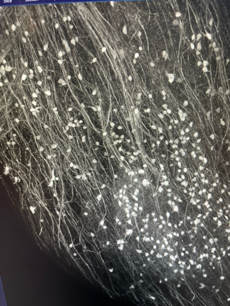
🧪 Tyrosine Hydroxylase is often the first probe labs try in order to validate their #clearing and labeling for #lightsheet #microscopy. It is robust, easy to identify and frankly, pretty. Need assistance with lightsheet? We are here to help at #Vanderbilt University. VanderbiltNL.com
November 22, 2024 at 2:56 AM
🧪 Tyrosine Hydroxylase is often the first probe labs try in order to validate their #clearing and labeling for #lightsheet #microscopy. It is robust, easy to identify and frankly, pretty. Need assistance with lightsheet? We are here to help at #Vanderbilt University. VanderbiltNL.com
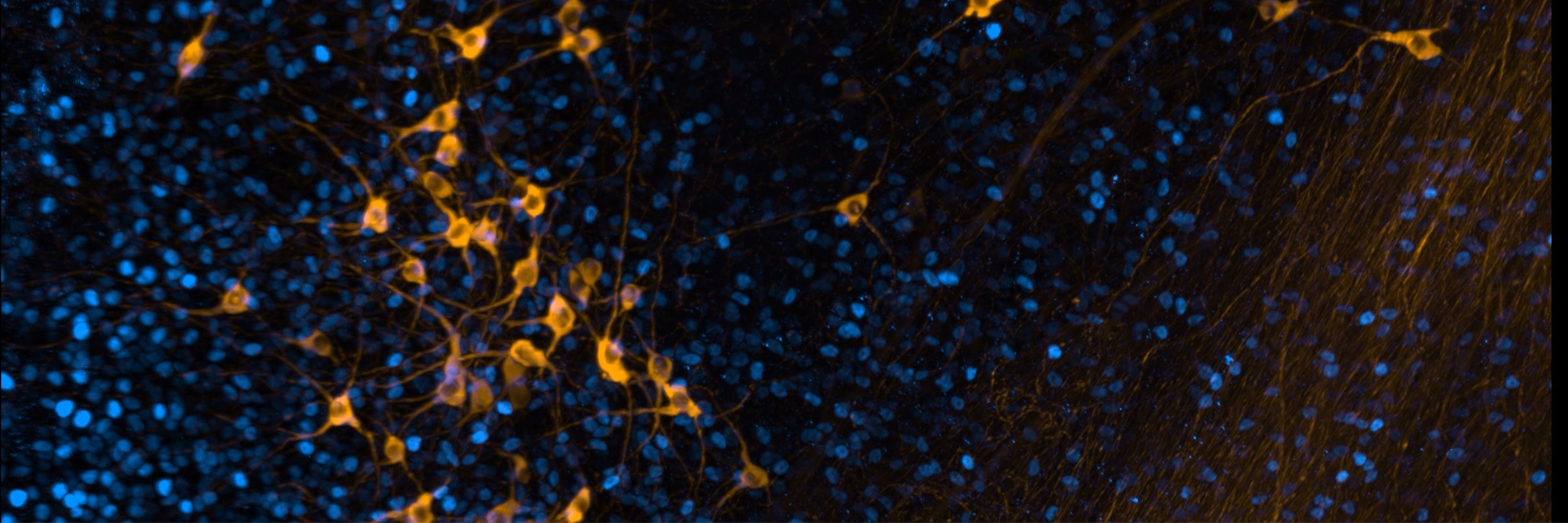
#Vanderbilt Neurovisualization lab specializes in #lightsheet #Microscopy. 🧪 Here we see a wave of c-fos positive cells in blue and tyrosine hydroxylase in gold. If you need help with lightsheet let me know!
November 19, 2024 at 4:57 PM
#Vanderbilt Neurovisualization lab specializes in #lightsheet #Microscopy. 🧪 Here we see a wave of c-fos positive cells in blue and tyrosine hydroxylase in gold. If you need help with lightsheet let me know!
One more #science video for today. Check out this forest of #neurons in a cleared #mouse #brain imaged with #lightsheet #microscopy at #Vanderbilt
November 19, 2024 at 1:06 AM
One more #science video for today. Check out this forest of #neurons in a cleared #mouse #brain imaged with #lightsheet #microscopy at #Vanderbilt
GFAP expressing #astrocytes in orange, IBA1 positive microglia in blue. Images from unsectioned rat hemisphere. #lightsheet #microscopy at #Vanderbilt
November 19, 2024 at 12:43 AM
GFAP expressing #astrocytes in orange, IBA1 positive microglia in blue. Images from unsectioned rat hemisphere. #lightsheet #microscopy at #Vanderbilt
Let’s get some #microscopy on bluesky going. Blood vessels in mouse brain imaged with #lightsheet microscopy #Vanderbilt
November 19, 2024 at 12:38 AM
Let’s get some #microscopy on bluesky going. Blood vessels in mouse brain imaged with #lightsheet microscopy #Vanderbilt