


Alain Dagher
@alaindagher.bsky.social
Neurology and brain imaging.
ORCID: 0000-0002-0945-5779
ORCID: 0000-0002-0945-5779
I have really enjoyed this book.

October 16, 2025 at 6:39 PM
I have really enjoyed this book.
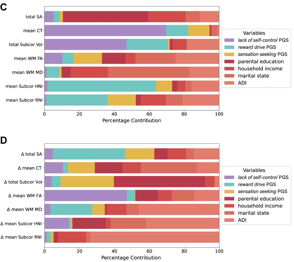
Finally, we found that each brain measure has a different ratio of genetic vs environmental influences. For example, cortical thickness was better captured by impulsivity PGSs, but surface area was more environmental.
June 16, 2025 at 6:23 PM
Finally, we found that each brain measure has a different ratio of genetic vs environmental influences. For example, cortical thickness was better captured by impulsivity PGSs, but surface area was more environmental.
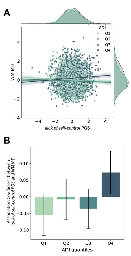
Next: how does environment come into play? We found that Area Deprivation Index modulates the relationship between lack of self-control PGS and white matter. Lower ADI amplified the effect of genetic risk on WM development.
June 16, 2025 at 6:23 PM
Next: how does environment come into play? We found that Area Deprivation Index modulates the relationship between lack of self-control PGS and white matter. Lower ADI amplified the effect of genetic risk on WM development.
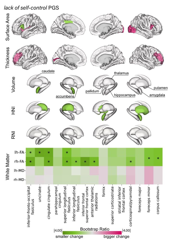
We also tested longitudinal changes in brain anatomy! Lack of self-control PGS was mostly related to slower development of white matter.
June 16, 2025 at 6:23 PM
We also tested longitudinal changes in brain anatomy! Lack of self-control PGS was mostly related to slower development of white matter.
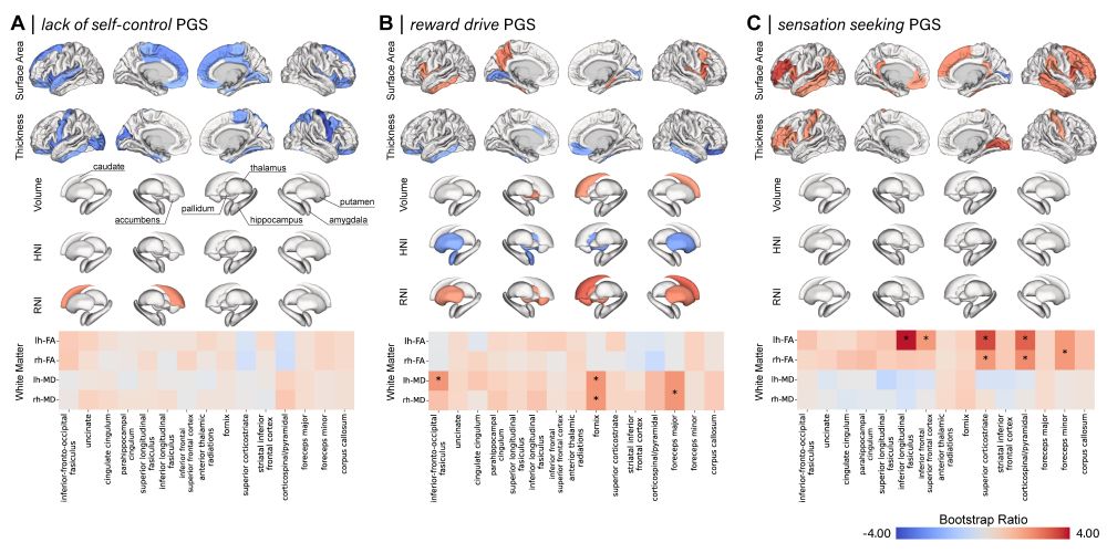
We then asked how the polygenic score for each latent factor manifested in the brain using ABCD (9-11 yrs, N>4,000). Lack of self-control: reduced prefrontal GM; reward drive: increased cellularity in subcortical nuclei; sensation seeking: increased surface area and white matter FA.
June 16, 2025 at 6:23 PM
We then asked how the polygenic score for each latent factor manifested in the brain using ABCD (9-11 yrs, N>4,000). Lack of self-control: reduced prefrontal GM; reward drive: increased cellularity in subcortical nuclei; sensation seeking: increased surface area and white matter FA.
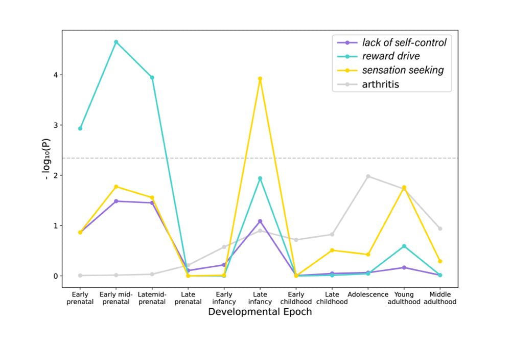
Gene-enrichment analysis showed that “reward drive” is preferentially associated with genes related to neurogenesis expressed at prenatal stage, while “sensation seeking” was associated with genes related to synaptic properties and expressed at late infancy.
June 16, 2025 at 6:23 PM
Gene-enrichment analysis showed that “reward drive” is preferentially associated with genes related to neurogenesis expressed at prenatal stage, while “sensation seeking” was associated with genes related to synaptic properties and expressed at late infancy.
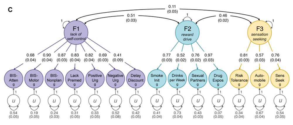
Applying gSEM to 17 GWASs, we discovered that different measures of impulsivity and risk-taking could be grouped into three factors based on their genetic architecture. We named them “lack of self-control,” “reward drive,” and “sensation seeking.”
June 16, 2025 at 6:23 PM
Applying gSEM to 17 GWASs, we discovered that different measures of impulsivity and risk-taking could be grouped into three factors based on their genetic architecture. We named them “lack of self-control,” “reward drive,” and “sensation seeking.”
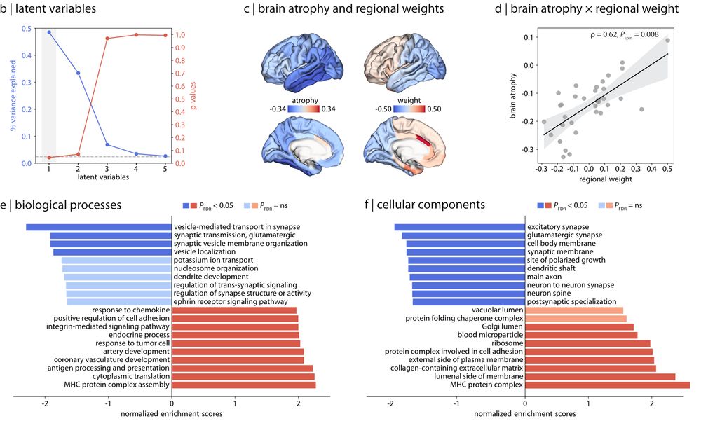
Imaging transcriptomics with the @alleninstitute.org Human Brain atlas identified the biological processes related to brain atrophy in PD, mostly related to excitatory synapses
May 28, 2025 at 5:12 PM
Imaging transcriptomics with the @alleninstitute.org Human Brain atlas identified the biological processes related to brain atrophy in PD, mostly related to excitatory synapses
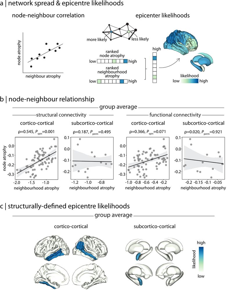
To test the prion-spread theory of PD, we found that cortical atrophy in any region was proportional to atrophy of its connected regions. We further identified the likeliest epicentres of disease initiation as the temporal lobe and amygdala, consistent with the brain-first route of propagation
May 28, 2025 at 5:12 PM
To test the prion-spread theory of PD, we found that cortical atrophy in any region was proportional to atrophy of its connected regions. We further identified the likeliest epicentres of disease initiation as the temporal lobe and amygdala, consistent with the brain-first route of propagation
Widespread cortical thinning was the most prominent finding when correlating MRI measures to disease stage and duration, and global cognitive score. It is thought that cortical thinning represents loss of synapses and neuropil.
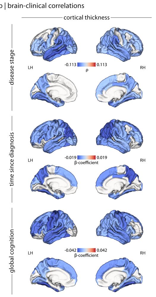
May 28, 2025 at 5:12 PM
Widespread cortical thinning was the most prominent finding when correlating MRI measures to disease stage and duration, and global cognitive score. It is thought that cortical thinning represents loss of synapses and neuropil.
New preprint. @ndrewvo.bsky.social and @enigmabrains.bsky.social analyzed MRI of > 3,000 people with Parkinson’s disease. W-score method to compare PD to matched controls confirmed extensive loss of cortical thickness, surface area and subcortical volumes in PD, even in early stages
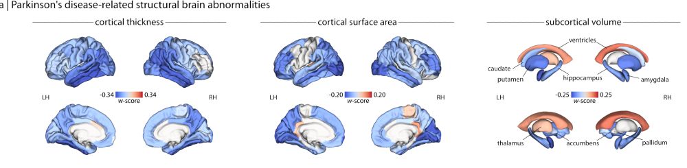
May 28, 2025 at 5:12 PM
New preprint. @ndrewvo.bsky.social and @enigmabrains.bsky.social analyzed MRI of > 3,000 people with Parkinson’s disease. W-score method to compare PD to matched controls confirmed extensive loss of cortical thickness, surface area and subcortical volumes in PD, even in early stages
6. We use BrainSpan to plot the developmental trajectory of energy genes. The main energy pathways follow brain (neocortical) size. Two notable exceptions: PPP highest during fetal development when tissue biogenesis is at its peak, and Ketone Body utilization peaks in infancy during breast feeding


March 18, 2025 at 9:48 PM
6. We use BrainSpan to plot the developmental trajectory of energy genes. The main energy pathways follow brain (neocortical) size. Two notable exceptions: PPP highest during fetal development when tissue biogenesis is at its peak, and Ketone Body utilization peaks in infancy during breast feeding
5. We focus on the visual system and identify a distinction between the dorsal and ventral stream. The dorsal stream encodes high- temporal frequency stimuli, and shows greater expression of Glycolysis, TCA, OxPhos and lactate pathways - components of rapid fuel supply from astrocytes to neurons .


March 18, 2025 at 9:48 PM
5. We focus on the visual system and identify a distinction between the dorsal and ventral stream. The dorsal stream encodes high- temporal frequency stimuli, and shows greater expression of Glycolysis, TCA, OxPhos and lactate pathways - components of rapid fuel supply from astrocytes to neurons .
4. Another distinction is between areas with large projection neurons (e.g. Betz cells in M1) with greater Glycolysis and OxPhos and sensory areas with dense inputs but short outputs – with higher expression of the PPP and TCA [Glycolysis/OxPhos aligns with the second functional gradient]

March 18, 2025 at 9:48 PM
4. Another distinction is between areas with large projection neurons (e.g. Betz cells in M1) with greater Glycolysis and OxPhos and sensory areas with dense inputs but short outputs – with higher expression of the PPP and TCA [Glycolysis/OxPhos aligns with the second functional gradient]
3. A consistent distinction emerges between glucose as fuel (glycolysis/OxPhos) and glucose as substrate for tissue biosynthesis and antioxidant defense (mainly PPP).

March 18, 2025 at 9:48 PM
3. A consistent distinction emerges between glucose as fuel (glycolysis/OxPhos) and glucose as substrate for tissue biosynthesis and antioxidant defense (mainly PPP).
New: Energy use shapes brain organization and development. We use gene expression maps from the @alleninstitute.bsky.social Human Brain Atlas to map the main energy metabolism pathways in the brain. Led by @moohebatpe.bsky.social @misicbata.bsky.social @goliashf.bsky.social and Justine Hansen 🧵

March 18, 2025 at 9:48 PM
New: Energy use shapes brain organization and development. We use gene expression maps from the @alleninstitute.bsky.social Human Brain Atlas to map the main energy metabolism pathways in the brain. Led by @moohebatpe.bsky.social @misicbata.bsky.social @goliashf.bsky.social and Justine Hansen 🧵
I am really enjoying this Naxos series of Russian Piano Trios - I only know some of this music - but it's all good. Highly recommend. music.apple.com/ca/album/his...


February 12, 2025 at 10:38 PM
I am really enjoying this Naxos series of Russian Piano Trios - I only know some of this music - but it's all good. Highly recommend. music.apple.com/ca/album/his...
Some say that BMI is an imperfect measure of metabolic risk. One reason is that some athletes can have BMI in the risky range despite very low adiposity. So I made a chart to help out.

January 15, 2025 at 4:36 PM
Some say that BMI is an imperfect measure of metabolic risk. One reason is that some athletes can have BMI in the risky range despite very low adiposity. So I made a chart to help out.
Agreed! It's super-interesting.
Did you know the Broca tried to measure focal brain activity with small temperature probes mounted on the scalp (Broca P. Sur les temperatures morbides locales, 1879) ? Later, Penfield used temp probes to directly measure cortical activation during surgery (see fig.)
Did you know the Broca tried to measure focal brain activity with small temperature probes mounted on the scalp (Broca P. Sur les temperatures morbides locales, 1879) ? Later, Penfield used temp probes to directly measure cortical activation during surgery (see fig.)

January 10, 2025 at 3:45 PM
Agreed! It's super-interesting.
Did you know the Broca tried to measure focal brain activity with small temperature probes mounted on the scalp (Broca P. Sur les temperatures morbides locales, 1879) ? Later, Penfield used temp probes to directly measure cortical activation during surgery (see fig.)
Did you know the Broca tried to measure focal brain activity with small temperature probes mounted on the scalp (Broca P. Sur les temperatures morbides locales, 1879) ? Later, Penfield used temp probes to directly measure cortical activation during surgery (see fig.)
That wasn't Mosso's first "brain imaging" experiment - also it wouldn't work (total blood volume shouldn't change due to brain activation).
Years earlier, however, he did show increases in DLPFC cerebral blood flow in a patient with a skull defect - see attached figure (courtesy of Albert Gjedde).
Years earlier, however, he did show increases in DLPFC cerebral blood flow in a patient with a skull defect - see attached figure (courtesy of Albert Gjedde).

January 9, 2025 at 4:16 PM
That wasn't Mosso's first "brain imaging" experiment - also it wouldn't work (total blood volume shouldn't change due to brain activation).
Years earlier, however, he did show increases in DLPFC cerebral blood flow in a patient with a skull defect - see attached figure (courtesy of Albert Gjedde).
Years earlier, however, he did show increases in DLPFC cerebral blood flow in a patient with a skull defect - see attached figure (courtesy of Albert Gjedde).

I like Stan Dehaene's take here. The hard problem of consciousness won't be solved, it will simply vanish like the 19th century concept of vitalism. www.softouch.on.ca/kb/data/Cons...

December 11, 2024 at 3:19 PM
I like Stan Dehaene's take here. The hard problem of consciousness won't be solved, it will simply vanish like the 19th century concept of vitalism. www.softouch.on.ca/kb/data/Cons...
Here's the Times:
November 26, 2024 at 12:43 AM
Here's the Times:
McConnell Brain Imaging Centre director Julien Doyon introduces the 40th anniversary celebrations.

November 19, 2024 at 2:38 PM
McConnell Brain Imaging Centre director Julien Doyon introduces the 40th anniversary celebrations.
"Devotional" by Celesti Kozub, first shown at OHBM in Rome, is back at the MNI.

November 18, 2024 at 11:02 PM
"Devotional" by Celesti Kozub, first shown at OHBM in Rome, is back at the MNI.